الأعضاء الأثرية في البشر
| د.إيهاب عبد الرحيم محمد ساهم بشكل رئيسي في تحرير هذا المقال
|
In the context of human evolution, human vestigiality involves those traits (such as organs or behaviors) occurring in humans that have lost all or most of their original function through evolution. Although structures called vestigial often appear functionless, a vestigial structure may retain lesser functions or develop minor new ones. In some cases, structures once identified as vestigial simply had an unrecognized function. Vestigial organs are sometimes called rudimentary organs.[1]
The examples of human vestigiality are numerous, including the anatomical (such as the human tailbone, wisdom teeth, and inside corner of the eye), the behavioral (goose bumps and palmar grasp reflex), and molecular (pseudogenes). Many human characteristics are also vestigial in other primates and related animals.
التاريخ
في الفصل الأول من كتابه عن نشوء الإنسان، ذكر داروين نحو عشرة صفات تشريحية وصفها بسعادة على أنها "غير ذات فائدة، أو تقترب من ذلك، وبالتالي فلم تعد عرضة للانتخاب الطبيعي." وقد تضمنت قائمة داروين شعر الجسم، وضرس العقل، والعصعص- وهي الخصائص التي استخدمها كدليل على صحة نظريته القائلة بأن البشر لم ينحدروا من سلالة أنصاف الآلهة، بل من سلسلة طويلة من المخلوقات المكسوة بالشعر، والتي تقتات على النباتات، والتي كان لها ذيل.
ولن نناقش هنا نظرية التطور لداروين، فيكفينا أن نذكر الآية الكريمة "ولقد خلقنا الإنسان في أحسن تقويم"، لنعلم أن الإنسان خُلق إنسانا ولم ينحدر عن القرود والنسانيس، بل سنناقش تلك الأعضاء التي لا يزال العلم عاجزا عن معرفة وظيفتها بالنسبة لأجسادنا البشرية، والتي لابد أن يأتي يوم ونعلم الحكمة من وجودها ... "وما أوتيتم من العلم إلا قليلا". كانت قائمة داروين بعيدة كل البُعد عن أن تكون مكتملة – فأجسامنا مليئة بأجزاء يبدو أننا لا نحتاج إليها، أو لا نفهم سبب وجودها ، وسنحاول في هذا المقال تجميعها للتفكر في خلق الله وتأمل بديع صنعه في الجسم البشري.
وبرغم مرور أكثر من قرن وربع القرن على وفاة داروين، لا يزال العلم عاجزا عن تقديم تفسير متكامل لسبب بقاء صفة تشريحية "متقادمة" ، ضمن المجموع الوراثي البشري، في حين اختفت صفات أخرى. وقد أظهرت أبحاث الجينوم الحديثة أن الدنا DNA البشري يحمل جينات معطوبة لأشياء قد تبدو مفيدة لو بقيت، مثل مستقبلات الرائحة التي تمنح حاسة شم قوية مثل تلك التي تتمتع بها الكلاب، أو إنزيمات كانت تتيح للبشر يوما تصنيع مخزوناتهم من فيتامين C. وقد تختفي صفات أخرى من البنية التشريحية للبشر خلال مئات أو آلاف السنين، لذلك فلننتهز الفرصة ونستعرضها الآن قبل اختفائها...
الأعضاء الأثرية
الجيوب الأنفية
 مقالة مفصلة: جيوب أنفية
قد تكون الجيوب الأنفية للبشر القدامى مبطنة بمستقبلات للرائحة تمنحهم حاسة شم قوية للغاية، مما ساعدهم على البقاء على قيد الحياة. أما الآن، فلا أحد يعلم على وجه التحديد لماذا نمتلك هذه الفراغات المبطنة بغشاء مخاطي ، والتي تسبب لنا كثيرا من المتاعب في كثير من الأحيان، اللهم إلا لجعل الرأس أخف، ولترطيب وتدفئة الهواء الذي نتنفسه. عند النظر إلى قطاع عرضي في الجمجمة البشرية، سنجد أربعة مجموعات من الجيوب الأنفية؛ الجبهية frontal – وتوجد في مقدم الرأس ، أو الجبهة ؛ والفكية العلوية maxillary – وتوجد تحت الخدين؛ والغربالية ethmoid والوتدية sphenoid- وتوجد خلف الأنف. وفي الحيوانات التي تتمتع بحاسة شم قوية، تبطن الجيوب الأنفية في معظمها بنسيج شمي.
العضو الميكعي الأنفي
 مقالة مفصلة: العضو الميكعي الأنفي
العضو الميكعي الأنفي Vomeronasal organ، هو حفرة صغيرة على كل من جانبي الحاجز الأنفي، وهو مبطن بمستقبلات كيميائية لا تعمل، أو غير معروفة الوظيفة، وربما كانت في أجدادنا تضطلع بوظيفة اكتشاف الفيرومونات pheromones- وهي مواد كيميائية تنتجها الحيوانات الثديية من الفئران إلى الأفيال،والتي تساعد على نقل المعلومات بشأن الحالة الاجتماعية ومتى يصبح الحيوان مستعدا للتناسل. ويفرز البشر مواد تشبه الفيرومونات لكن ليس بالكيفية التي عليها بقية الثدييات. وبينما يستخدم البشر عيونهم لتحديد شريك العمر وصفاته الأخرى، تستخدم بقية الثدييات "الأنف الثاني" أو العضو الميكعي الأنفي؛ فقد اكتشف الباحثون في الولايات المتحدة أن الفئران عندما تقرر التزاوج فإنها تستخدم عضوا أساسيا غير متوقع هو عبارة عن أنف ثان تستطيع من خلاله أن تميز بين جنس شريك العمر وحالته الاجتماعية ومدى توافر مشاعر عاطفية متبادلة.إن الأنف الأساسي للفأر ربما يخبره بمكان الطعام، لكن عضوا منفصلا –هو العضو الميكعي الأنفي- يفتح عالما مختلفا من الإدراك الحسي، ويشبه صندوقا أسود في المخ، وهو عبارة عن هيكل أنبوبي صغير جدا يشبه اللسان اكتشف في قاعدة الأنف الأساسية. وذكرت الأبحاث الحديثة وصفا لكيف أن الخلايا العصبية المحددة للفيرومونات في الأنف الثاني تعدل جهاز استقبالها ليتعرف على جنس الفأر الثاني ونوعه. فهل لهذا دور في البشر أيضا؟ ربما... فقد أفادت دراسة يابانية أن الفيرومون الموجود في عَرَق الرجال قد يكون له أثر فعال على النساء جسديا ونفسيا. فقد أشارت الدراسة لأن تعرض انف المرأة لرائحة العَرَق الذكري والذي يحتوي على الفيرومون يؤدي إلى تحسن جذري في مزاج المرأة ويساعد في إطلاق هرمونات في جسم المرأة تؤدي إلى تنظيم الدورة الشهرية عندها.
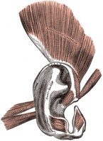
العضلات الخارجية للأذن
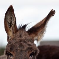
تسمح هذه العضلات الثلاث لحيوانات مثل الكلاب والأرانب تحريك آذانها بحرّية ، لكن وظيفتها في البشر غير معلومة، بيد أنه موجودة لدى جميع البشر، ويمكن لأي إنسان أن يتعلم تحريكها بنوع من التدريب.
الضلع الرقبي
 مقالة مفصلة: الضلع الرقبي
هناك زوج من الضلوع الرقبية الشبيهة بتلك الموجودة في الزواحف ، في أقل من 1% من البشر. وبالإضافة إلى أن وظيفتها غير معروفة، فهي تسبب العديد من المشكلات العصبية والشريانية في الأشخاص الذين يمتلكونها.
الجفن الثالث
 مقالة مفصلة: غشاء راف
تمتلك الطيور وبعض الثدييات غشاء إضافياً، الجفن الثالث أو الغشاء الراف، تحت الجفن لحماية العين وتنظيفها من الأتربة، لكن البشر لا يمتلكون منه سوى طية صغيرة عند الركن الداخلي لكل من العينين تسمى الموق.
نقطة داروين
 مقالة مفصلة: نتوء داروين
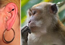
The ears of a macaque monkey and most other monkeys have far more developed muscles than those of humans, and therefore have the capability to move their ears to better hear potential threats.[2] Humans and other primates such as the orangutan and chimpanzee however have ear muscles that are minimally developed and non-functional, yet still large enough to be identifiable.[3] A muscle attached to the ear that cannot move the ear, for whatever reason, can no longer be said to have any biological function. In humans there is variability in these muscles, such that some people are able to move their ears in various directions, and it can be possible for others to gain such movement by repeated trials.[3][4] In such primates, the inability to move the ear is compensated mainly by the ability to turn the head on a horizontal plane, an ability which is not common to most monkeys—a function once provided by one structure is now replaced by another.[5]
The outer structure of the ear also shows some vestigial features, such as the node or point on the helix of the ear known as Darwin's tubercle which is found in around 10% of the population.
العين
The plica semilunaris is a small fold of tissue on the inside corner of the eye. It is the vestigial remnant of the nictitating membrane, i.e., third eyelid, an organ that is fully functional in some other species of mammals.[6] Its associated muscles are also vestigial.[3] Only one species of primate, the Calabar angwantibo, is known to have a functioning nictitating membrane.[7]
The orbitalis muscle is a vestigial or rudimentary nonstriated muscle (smooth muscle) of the eye that crosses from the infraorbital groove and sphenomaxillary fissure and is intimately united with the periosteum of the orbit. It was described by Johannes Peter Müller and is often called Müller's muscle. The muscle forms an important part of the lateral orbital wall in some animals, but in humans it is not known to have any significant function.[8][9]
الجهاز التناسلي
Genitalia
In the internal genitalia of each human sex, there are some residual organs of mesonephric and paramesonephric ducts during embryonic development:
Human vestigial structures also include leftover embryological remnants that once served a function during development, such as the belly button, and analogous structures between biological sexes. For example, men are also born with two nipples, which are not known to serve a function compared to women.[10] In regards to genitourinary development, both internal and external genitalia of male and female fetuses have the ability to fully or partially form their analogous phenotype of the opposite biological sex if exposed to a lack/overabundance of androgens or the SRY gene during fetal development.[11][12] Examples of vestigial remnants of genitourinary development include the hymen, which is a membrane that surrounds or partially covers the external vaginal opening that derives from the sinus tubercle during fetal development and is homologous to the male seminal colliculus.[13] Some researchers[من؟] have hypothesized that the persistence of the hymen may be to provide temporary protection from infection, as it separates the vaginal lumen from the urogenital sinus cavity during development.[14] Other examples include the glans penis and the clitoris, the labia minora and the ventral penis, and the ovarian follicles and the seminiferous tubules.[13]
In modern times, there is controversy regarding whether the foreskin is a vital or vestigial structure.[15] In 1949, British physician Douglas Gairdner noted that the foreskin plays an important protective role in newborns. He wrote, "It is often stated that the prepuce is a vestigial structure devoid of function ... However, it seems to be no accident that during the years when the child is incontinent the glans is completely clothed by the prepuce, for, deprived of this protection, the glans becomes susceptible to injury from contact with sodden clothes or napkin."[15] During the physical act of sex, the foreskin reduces friction, which can reduce the need for additional sources of lubrication.[15] "Some medical researchers, however, claim circumcised men enjoy sex just fine and that, in view of recent research on HIV transmission, the foreskin causes more trouble than it's worth."[15] However, recent Canadian studies on Circumcision & HIV risk have thrown this conclusion into question[16] The area of the outer foreskin measures between 7 and 100 cm2,[17] and the inner foreskin measures between 18 and 68 cm2,[18] which is a wide range. Regarding vestigial structures, Charles Darwin wrote, "An organ, when rendered useless, may well be variable, for its variations cannot be checked by natural selection."[19] Charles Darwin speculated that the sensitivity of the foreskin to fine touch might have served as an "early warning system" in our naked ancestors while it protected the glans from the intrusion of biting insects and parasites.[19]
الجهاز العضلي
A number of muscles in the human body are thought to be vestigial, either by virtue of being greatly reduced in size compared to homologous muscles in other species, by having become principally tendonous, or by being highly variable in their frequency within or between populations.
Head
The occipitalis minor is a muscle in the back of the head which normally joins to the auricular muscles of the ear. This muscle is very sporadic in frequency—always present in Malays, present in 56% of Africans, 50% of Japanese, and 36% of Europeans, and nonexistent in the Khoikhoi people of southwestern Africa and in Melanesians.[20] Other small muscles in the head associated with the occipital region and the post-auricular muscle complex are often variable in their frequency.[21]
The platysma, a quadrangular (four sides) muscle in a sheet-like configuration, is a vestigial remnant of the panniculous carnosus of animals. In horses, it is the muscle that allows it to flick a fly off its back.
Face
In many lower animals, the upper lip and sinus area is associated with whiskers or vibrissae which serve a sensory function. In humans, these whiskers do not exist but there are still sporadic cases where elements of the associated vibrissal capsular muscles or sinus hair muscles can be found. Based on histological studies of the upper lips of 20 cadavers, Tamatsu et al. found that structures resembling such muscles were present in 35% (7/20) of their specimens.[22]
Arm
The palmaris longus muscle is seen as a small tendon between the flexor carpi radialis and the flexor carpi ulnaris, although it is not always present. The muscle is absent in about 14% of the population, however this varies greatly with ethnicity. It is believed that this muscle actively participated in the arboreal locomotion of primates, but currently has no function, because it does not provide more grip strength.[23] One study has shown the prevalence of palmaris longus agenesis in 500 Indian patients to be 17.2% (8% bilateral and 9.2% unilateral).[24] The palmaris is a popular source of tendon material for grafts and this has prompted studies which have shown the absence of the palmaris does not have any appreciable effect on grip strength.[25]
The levator claviculae muscle in the posterior triangle of the neck is a supernumerary muscle present in only 2–3% of all people[26] but nearly always present in most mammalian species, including gibbons and orangutans.[27]
Torso
The pyramidalis muscle of the abdomen is a small and triangular muscle, anterior to the rectus abdominis, and contained in the rectus sheath. It is absent in 20% of humans and when absent, the lower end of the rectus then becomes proportionately increased in size. Anatomical studies suggest that the forces generated by the pyramidalis muscles are relatively small.[28]
The latissimus dorsi muscle of the back has several sporadic variations. One particular variant is the existence of the dorsoepitrochlearis or latissimocondyloideus muscle which is a muscle passing from the tendon of the latissimus dorsi to the long head of the triceps brachii. It is notable due to its well developed character in other apes and monkeys, where it is an important climbing muscle, namely the dorsoepitrochlearis brachii.[29][30] This muscle is found in ≈5% of humans.[31]
Leg
The plantaris muscle is composed of a thin muscle belly and a long thin tendon. The muscle belly is approximately 5–10 سنتيمتر (2–4 بوصات) long, and is absent in 7–10% of the human population. It has some weak functionality in moving the knee and ankle but is generally considered redundant and is often used as a source of tendon for grafts. The long, thin tendon of the plantaris is humorously called "the freshman's nerve", as it is often mistaken for a nerve by new medical students.
Tongue
Another example of human vestigiality occurs in the tongue, specifically the chondroglossus muscle. In a morphological study of 100 Japanese cadavers, it was found that 86% of fibers identified were solid and bundled in the appropriate way to facilitate speech and mastication. The other 14% of fibers were short, thin and sparse – nearly useless, and thus concluded to be of vestigial origin.[32]
الثديان
Extra nipples or breasts sometimes appear along the mammary lines of humans, appearing as a remnant of mammalian ancestors who possessed more than two nipples or breasts.[33][34] One recent report demonstrated that all healthy young men and women who participated in an anatomic study of the front surface of the body exhibited 8 pairs of focal fat mounds running along the embryological mammary ridges from axillae to the upper inner thighs. These were always located in the same relative anatomic sites – analogous to the loci of breasts in other placental mammals – and often had nipple-like moles or extra hairs located atop the mounds. Therefore, focal fatty prominences on the fronts of human torsos likely represent chains of vestigial breasts composed of primordial breast fat.[35]
تتكون القنوات الحاملة للّبن قبل فترة طويلة من قيام هرمون الذكورة (التستوستيرون) بإحداث التمايز الجنسي في الجنين إلى ذكر أو أنثى؛ لكن الذكور من البشر يمتلكون نسيجا ثدييا يمكن تحريضه على إنتاج اللبن، كما يتضخم الثدي في الذكور في بعض الاضطرابات الهرمونية ، وهو ما يسمى بتثدي الرجل gynecomastia.
العضلة تحت الترقوية
 مقالة مفصلة: عضلة تحت ترقوية
العضلة تحت الترقوية، هي عضلة صغيرة تمتد تحت الكتف من الضلع الأول إلى الترقوة؛ وهي مفيدة للحيوانات التي تسير على أربع ؛ أما البشر فيمتلك بعضهم واحدة، وبعضهم لا يمتلكها على الإطلاق، وقليل هم من يمتلكون زوجا من هذه العضلات – واحدة على كل من الجانبين.
العضلة الراحية الطويلة
 مقالة مفصلة: العضلة الراحية الطويلة
تمتد هذه العضلة الطويلة النحيلة من المرفق إلى الرسغ ، وتوجد لدى 89% من البشر المعاصرين؛ وربما كانت مفيدة لدى أجدادنا من البشر القدامى في تسلق الأشجار. ويستخدم الجراحون تلك العضلة في جراحات التجميل عند الحاجة لزرع عضلة من جسم نفس المريض، إذ لا يؤدي استئصال العضلة الراحية إلى أي تأثير على وظيفة اليد.
القشعريرة
 مقالات مفصلة: قشعريرة
- عضلة مقفة للشعرة
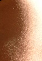
العضلة المقفة للشعر Arrector pili muscle، هي حزم من الألياف العضلية الملساء التي تمكّن الحيوانات من نفش فروها بغرض الوقاية من البرد أو لتخويف الحيوانات الأخرى. ولا يزال البشر يحتفظون بتلك القدرة ، كما يتضح عندما نصاب بالقشعريرة (جلد الإوز)، لكن الواضح أنهم لا يمتلكون معظم الفرو الموجود لدى الحيوانات الأخرى.
The formation of goose bumps in humans under stress is a vestigial reflex; a possible function in the distant evolutionary ancestors of humanity was to raise the body's hair, making the ancestor appear larger and scaring off predators.[36][37] Raising the hair is also used to trap an extra layer of air, keeping an animal warm.[37] Due to the diminished amount of hair in humans, the reflex formation of goose bumps when cold is also vestigial.[37]
الزائدة الدودية
 مقالة مفصلة: زائدة دودية
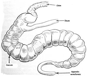
كانت هذه القناة العضلية الصغيرة المتصلة بالأمعاء الغليظة مفيدة عندما كان طعام أجدادنا من البشر جامعي الثمار يتكون في معظمه من مواد نباتية أكثر من احتوائه على البروتينات الحيوانية المصدر، إذ تعمل الزائدة الدودية كمنطقة خاصة لهضم السليولوز ، كما تنتج كذلك بعض كريات الدم البيضاء. أما الآن ، فبالإضافة إلى أننا لا نعلم وظيفتها على وجه التحديد، فهي تسبب مشكلات كبيرة عند التهابها ، فيجري استئصالها جراحيا ، وهي عملية تجرى أكثر من 300,000 مرة سنويا في الولايات المتحدة وحدها.
In modern humans, the appendix is sometimes believed to be a vestige of a redundant organ that in ancestral species had digestive functions, much as it still does in extant species in which intestinal flora hydrolyze cellulose and similar indigestible plant materials.[3] This view has changed over the past decades,[38] with research suggesting that the appendix may serve an important purpose. In particular, it may serve as a reservoir for beneficial gut bacteria.
Some herbivorous animals, such as rabbits, have a terminal vermiform appendix and cecum that apparently bear patches of tissue with immune functions and may also be important in maintaining the composition of intestinal flora. It does not however seem to have much digestive function, if any, and is not present in all herbivores, even those with large caeca.[39] As shown in the accompanying pictures however, the human appendix typically is about comparable to that of the rabbit's in size, though the caecum is reduced to a single bulge where the ileum empties into the colon.[40] Some carnivorous animals may have appendices too, but seldom have more than vestigial caeca.[41] In line with the possibility of vestigial organs developing new functions, some research suggests that the appendix may guard against the loss of symbiotic bacteria that aid in digestion, though that is unlikely to be a novel function, given the presence of vermiform appendices in many herbivores.[42][43] Intestinal bacterial populations entrenched in the appendix may support quick re-establishment of the flora of the large intestine after an illness, poisoning, or after an antibiotic treatment depletes or otherwise causes harmful changes to the bacterial population of the colon.[44]
A 2013 study, however, refutes the idea of an inverse relationship between cecum size and appendix size and presence. It is widely present in euarchontoglires (a superorder of mammals that includes rodents, lagomorphs and primates) and has also evolved independently in the diprotodont marsupials, monotremes, and is highly diverse in size and shape which could suggest it is not vestigial. Researchers deduce that the appendix has the ability to protect good bacteria in the gut. That way, when the gut is affected by a bout of diarrhea or other illness that cleans out the intestines, the good bacteria in the appendix can repopulate the digestive system and keep the person healthy.[45]
الفواق
It has been proposed that the hiccup is an evolutionary remnant of earlier amphibian respiration.[46] Amphibians such as tadpoles gulp air and water across their gills via a rather simple motor reflex akin to mammalian hiccuping. The motor pathways that enable hiccuping form early during fetal development, before the motor pathways that enable normal lung ventilation form. Additionally, hiccups and amphibian gulping are inhibited by elevated COقالب:Sub2 and may be stopped by GABAB receptor agonists, illustrating a possible shared physiology and evolutionary heritage. These proposals may explain why premature infants spend 2.5% of their time hiccuping, possibly gulping like amphibians, as their lungs are not yet fully formed. Fetal intrauterine hiccups are of two types. The physiological type occurs before 28 weeks after conception and tend to last five to ten minutes. These hiccups are part of fetal development and are associated with the myelination of the phrenic nerve, which primarily controls the thoracic diaphragm. The phylogeny hypothesis explains how the hiccup reflex might have evolved, and if there is not an explanation, it may explain hiccups as an evolutionary remnant, held-over from our amphibious ancestors. This hypothesis has been questioned because of the existence of the afferent loop of the reflex, the fact that it does not explain the reason for glottic closure, and because the very short contraction of the hiccup is unlikely to have a significant strengthening effect on the slow-twitch muscles of respiration.[بحاجة لمصدر]
ضروس العقل
 مقالة مفصلة: ضرس العقل
Wisdom teeth are vestigial third molars that human ancestors used to help in grinding down plant tissue. The common postulation is that the skulls of human ancestors had larger jaws with more teeth, which were possibly used to help chew down foliage to compensate for a lack of ability to efficiently digest the cellulose that makes up a plant cell wall. As human diets changed, smaller jaws were naturally selected, yet the third molars, or "wisdom teeth", still commonly develop in human mouths.[47] In modern human populations, wisdom teeth have become useless and often present harmful complications to the extent that surgical procedures are frequently performed to remove them.
Agenesis (failure to develop) of wisdom teeth in human populations ranges from zero in Tasmanian Aboriginals to nearly 100% in indigenous Mexicans.[48] The difference is related to the PAX9 gene (and perhaps other genes).[49]
شعر الجسم
 مقالة مفصلة: شعر (تشريح)
يساعد الحاجبان على وقاية العينين من تساقط العرق فيهما، وقد يلعب شعر الوجه في الذكور دوراً في الانتقاء الجنسي؛ لكنه من الواضح أن معظم الشعر المتبقي على أجساد البشر لا يلعب دوراً وظيفياً معروفاً.
العضلة الأخمصية
 مقالة مفصلة: العضلة الأخمصية
كثيرا ما يتم الخلط بين العضلة الأخمصية plantaris muscle- عند وجودها- وبين أحد الأعصاب في درس التشريح من قبل الطلاب المستجدين في كليات الطب. وهذه العضلة مفيدة في الرئيسات من غير البشر في قبض أرجلها للإمساك بفروع الأشجار، وهي غير موجودة أصلا لدى 9% من البشر المعاصرين.
الضلع الثالث عشر
يمتلك أقرب الرئيسات شبها بالإنسان – الشمبانزي والغوريلا- زوجاً إضافياً من الضلوع؛ أما نحن فمعظمنا يمتلك اثنا عشر زوجاً من الضلوع، لكن 8% من البشر البالغين يمتلكون زوجاً إضافياً من الضلوع التي لا نعلم لها وظيفة حتى الآن.
الرحم الذكري
هناك بقايا عضو تناسلي أنثوي رديم يتدلى من غدة البروستاتة في الذكور.
الإصبع الصغير للقدم
تحتاج القردة الدنيا لجميع أصابع أقدامها للقبض والتعلق بالأغصان والتنقل بينها ، لكن البشر يحتاجون إلى الإصبع الكبير للقدم بصورة أساسية للمحافظة على توازنهم عند الوقوف في الوضع المنتصب.
الأسهر الأنثوي
 مقالة مفصلة: الأسهر
إن العضو الذي يتحول إلى القنوات الناقلة للمني في الذكور يتحول في الإناث إلى المباض، وهو تجمّع من القنوات المسدودة التي لا وظيفة معروفة لها، ويقع قرب المبيض.
العضلة الهرمية
 مقالة مفصلة: العضلة الهرمية
العضلة الهرمية pyramidalis muscle، يفتقر أكثر من 20% منا لهذه العضلة الضئيلة المثلثة الشكل والشبيهة بالكيس، والملتصقة بعظم العانة ، وهي تشبه الكيس الموجود لدى الجرابيات، مثل الكنغر، لكنه فيها أكثر تطورا بكثير ويؤدي وظيفة تناسلية هامة.
العصعص
 مقالة مفصلة: عصعص
تمتلك أغلب الثدييات ذيلا تستخدمه في التوازن والاتصال، أما في البشر فلا يوجد سوى تلك الفقرات المندمجة، حيث لا يحتاج البشر لذيل إذ يمشون منتصبين، وهذا من تكريم الله تعالى لبني الإنسان، فتبارك الله أحسن الخالقين. ويتباين العصعص coccyx البشري كثيرا في الشكل والحجم، لكنه عموما يتكون من 3-5 فقرات مندمجة؛ وفي حالات نادرة ، يولد أطفال إما بدون عصعص على الإطلاق أو بذيل! وقد اقترح بعض الباحثين أن العصعص يساعد كموضع لارتكاز العضلات الصغيرة وفي دعم الأعضاء الحوضية، وذلك لأن استئصاله جراحيا ليس له تأثير واضح على صحة البشر.
الجين الكاذب
There are also vestigial molecular structures in humans, which are no longer in use but may indicate common ancestry with other species. One example of this is L-gulonolactone oxidase, a gene that is functional in most other mammals and produces an enzyme that synthesizes vitamin C.[50] In humans and other members of the suborder Haplorrhini, a mutation disabled the gene and made it unable to produce the enzyme. However, the remains of the gene are still present in the human genome as a vestigial genetic sequence called a pseudogene.[51]
انظر أيضاً
- عمى الألوان
- Deprecation
- قصر النظر
- Atavism
- Dewclaw
- Exaptation
- Human vestigiality
- Maladaptation
- العضلة الأخمصية
- Recessive refuge
- Spandrel (biology)
- الاستجابة الأثرية
المراجع
- ^ "Difference between rudimentary and vestigial organ - Biology - Evolution - 11741123 | Meritnation.com". www.meritnation.com. Retrieved 2021-02-16.
- ^ Prof. A. Macalister, Annals and Magazine of Natural History, vol. vii., 1871, p. 342.
- ^ أ ب ت ث Darwin, Charles (1871). The Descent of Man, and Selection in Relation to Sex. John Murray: London.
- ^ Bair, J. H. (1901). "Development of voluntary control". Psychological Review. 8 (5): 474–510. doi:10.1037/h0074157. hdl:2027/mdp.39015070189314.
- ^ Mr. St. George Mivart, Elementary Anatomy, 1873, p. 396.
- ^ Owen, R. 1866–1868. Comparative Anatomy and Physiology of Vertebrates. London.[صفحة مطلوبة]
- ^ Montagna, W.; Machida, H.; Perkins, E. M. (1966). "The skin of primates. XXXIII. The skin of the angwantibo (Arctocebus calabarensis)". American Journal of Physical Anthropology. 25 (3): 277–90. doi:10.1002/ajpa.1330250307. PMID 5971502.
- ^ Toerien, M. J.; Gous, A. E. (1978). "The orbital muscle of Müller". South African Medical Journal. 53 (4): 139–41. PMID 653491.
- ^ Dutton, J.J., Atlas of Clinical and Surgical Orbital Anatomy, 2nd Edition, Elsevier, 2011. p.116-117.
- ^ "Breast Anatomy and Embryology". Essentials of Plastic Surgery (2015): 355–361
- ^ Hadjiathanasiou, C.G.; Brauner, R.; Lortat-Jacob, S.; Nivot, S.; Jaubert, F.; Fellous, M.; Nihoul-Fékété, C.; Rappaport, R. (1994). "True hermaphroditism: Genetic variants and clinical management". The Journal of Pediatrics. 125 (5): 738–744. doi:10.1016/S0022-3476(06)80172-1. PMID 7965425.
- ^ Eren, Erdal; Edgünlü, Tuba; Asut, Emre; Karakaş Çelik, Sevim (2016). "Homozygous Ala65Pro Mutation with V89L Polymorphism in SRD5A2 Deficiency". Journal of Clinical Research in Pediatric Endocrinology. 8 (2): 218–223. doi:10.4274/jcrpe.2495. PMC 5096479. PMID 26761946.
- ^ أ ب Healey, Andrew (2010). "Embryology of the Female Reproductive Tract". Imaging of Gynecological Disorders in Infants and Children. Medical Radiology. pp. 21–30. doi:10.1007/174_2010_128. ISBN 978-3-540-85601-6.
- ^ Basaran, Mustafa; Usal, Deniz; Aydemir, Cumhur (2009). "Hymen Sparing Surgery for Imperforate Hymen: Case Reports and Review of Literature". Journal of Pediatric and Adolescent Gynecology. 22 (4): e61–64. doi:10.1016/j.jpag.2008.03.009. PMID 19646660.
- ^ أ ب ت ث Collier, Roger (2011-11-22). "Vital or vestigial? The foreskin has its fans and foes". CMAJ (in الإنجليزية). 183 (17): 1963–1964. doi:10.1503/cmaj.109-4014. ISSN 0820-3946. PMC 3225416. PMID 22025652.
- ^ Nayan, Madhur; Hamilton, Robert J.; Juurlink, David N.; Austin, Peter C.; Jarvi, Keith A. (2022-02-01). "Circumcision and Risk of HIV among Males from Ontario, Canada". Journal of Urology. 207 (2): 424–430. doi:10.1097/JU.0000000000002234.
- ^ Kigozi G, Wawer M, Ssettuba A, et al. "Foreskin surface area and HIV acquisition in Rakai, Uganda (size matters)". AIDS. 2009; 23(16):2209–2213. 10.1097/QAD.0b013e328330eda8.
- ^ Werker PMN, Terng ASC, Kon M. "The prepuce free flap: dissection feasibility study and clinical application of a super-thin new flap". Plastic Reconstructive Surgery. 1998; 102(4):1075–1082. 10.1097/00006534-199809020-00024.
- ^ أ ب Darwin C. The Origin of Species by Means of Natural Selection. London, UK: John Murray; 1859.
- ^ Macalister, Alexander (1875). "Additional Observations on Muscular Anomalies in Human Anatomy. (Third Series) With a Catalogue of the Principal Muscular Variations Hitherto Published". The Transactions of the Royal Irish Academy. 25: 1–134. JSTOR 30079154.
- ^ Guerra, Aldo Benjamin; Metzinger, Stephen Eric; Metzinger, Rebecca Crawford; Xie, Chen; Xie, Yue; Rigby, Peter Lister; Naugle, Thomas (2004). "Variability of the Postauricular Muscle Complex". Archives of Facial Plastic Surgery. 6 (5): 342–7. doi:10.1001/archfaci.6.5.342. PMID 15381582.
- ^ Tamatsu, Yuichi; Tsukahara, Kazue; Hotta, Mitsuyuki; Shimada, Kazuyuki (2007). "Vestiges of vibrissal capsular muscles exist in the human upper lip". Clinical Anatomy. 20 (6): 628–31. doi:10.1002/ca.20497. PMID 17458869. S2CID 21055062.
- ^ Aversi-Ferreira, Roqueline A. G. M. F.; Bretas, Rafael Vieira; Maior, Rafael Souto; Davaasuren, Munkhzul; Paraguassú-Chaves, Carlos Alberto; Nishijo, Hisao; Aversi-Ferreira, Tales Alexandre (2014). "Morphometric and Statistical Analysis of the Palmaris Longus Muscle in Human and Non-Human Primates". BioMed Research International. 2014: 1–6. doi:10.1155/2014/178906. PMC 4016873. PMID 24860810.
- ^ Kapoor, Sudhir K.; Tiwari, Akshay; Kumar, Abhishek; Bhatia, Rajesh; Tantuway, Vinay; Kapoor, Saurabh (2008). "Clinical relevance of palmaris longus agenesis: Common anatomical aberration". Anatomical Science International. 83 (1): 45–8. doi:10.1111/j.1447-073X.2007.00199.x. PMID 18402087. S2CID 25324691.
- ^ Sebastin, S; Lim, A; Bee, W; Wong, T; Methil, B (2005). "Does the absence of the palmaris longus affect grip and pinch strength?". The Journal of Hand Surgery: Journal of the British Society for Surgery of the Hand. 30 (4): 406–8. doi:10.1016/j.jhsb.2005.03.011. PMID 15935531. S2CID 35394120.
- ^ Rubinstein, David; Escott, Edward J.; Hendrick, Laura L. (April 1999). "The prevalence and CT appearance of the levator claviculae muscle: a normal variant not to be mistaken for an abnormality" (PDF). AJNR Am J Neuroradiol. 20 (4): 583–6. PMC 7056035. PMID 10319965.
- ^ Loukas, M.; Sullivan, A.; Tubbs, R.S.; Shoja, M.M. (2008). "Levator claviculae: a case report and review of the literature". Folia Morphol. 67 (4): 307–310. PMID 19085875.
- ^ Lovering, Richard M.; Anderson, Larry D. (2008). "Architecture and fiber type of the pyramidalis muscle". Anatomical Science International. 83 (4): 294–7. doi:10.1111/j.1447-073X.2007.00226.x. PMC 3531545. PMID 19159363.
- ^ P., Haninec; R., Tomáš; R., Kaiser; R., Čihák (2009). "Development and clinical significance of the musculus dorsoepitrochlearis in men". Clinical Anatomy. 22 (4): 481–8. doi:10.1002/ca.20799. PMID 19373904. S2CID 221547558.
- ^ Edwards, William E., The Musculoskeletal Anatomy of the Thorax and Brachium of an Adult Female Chimpanzee,6571st Aeromedical Research Laboratory, New Mexico, 1965. http://www.dtic.mil/dtic/tr/fulltext/u2/462433.pdf
- ^ "Anatomy Atlases: Illustrated Encyclopedia of Human Anatomic Variation: Opus I: Muscular System: Alphabetical Listing of Muscles: L:Latissimus Dorsi".
- ^ Ogata, Shigemitsu; Mine, Kazuharu; Tamatsu, Yuichi; Shimada, Kazuyuki (2002). "Morphological study of the human chondroglossus muscle in Japanese". Annals of Anatomy - Anatomischer Anzeiger. 184 (5): 493–9. doi:10.1016/S0940-9602(02)80087-5. PMID 12392330.
- ^ Kajava, Y (1915). "The proportions of supernumerary nipples in the Finnish population". Duodecim. 1: 143–70.
- ^ Goyal, Tarang; Bakshi, SK; Varshney, Anupam (2012). "Seven nipples in a male: World′s second case report". Indian Journal of Human Genetics. 18 (3): 373–5. doi:10.4103/0971-6866.108051. PMC 3656534. PMID 23716953.
{{cite journal}}: CS1 maint: unflagged free DOI (link) - ^ Teplica, David; Kovich, Grant; Srock, Jamey; Whitaker, Robert; Jeffers, Eileen; Wagstaff, David A. (2021-10-14). "Newly Identified Gross Human Anatomy: Eight Paired Vestigial Breast Mounds Run along the Embryological Mammary Ridges in Lean Adults". Plastic and Reconstructive Surgery - Global Open (in الإنجليزية). 9 (10): e3863. doi:10.1097/GOX.0000000000003863. ISSN 2169-7574. PMC 8517303. PMID 34667697.
- ^ Darwin, Charles. (1872) The Expression of the Emotions in Man and Animals John Murray, London.[صفحة مطلوبة]
- ^ أ ب ت خطأ استشهاد: وسم
<ref>غير صحيح؛ لا نص تم توفيره للمراجع المسماةSpinney - ^ Kooij IA, Sahami S, Meijer SL, Buskens CJ, Te Velde AA (October 2016). "The immunology of the vermiform appendix: a review of the literature". Clinical and Experimental Immunology. 186 (1): 1–9. doi:10.1111/cei.12821. PMC 5011360. PMID 27271818.
- ^ Stevens, C. Edward; Hume, Ian (2004). Comparative Physiology of the Vertebrate Digestive System. Cambridge: Cambridge University Press. ISBN 978-0-521-61714-7.
- ^ خطأ استشهاد: وسم
<ref>غير صحيح؛ لا نص تم توفيره للمراجع المسماةWellsSOF - ^ Peter Robert Cheeke, Ellen S. Dierenfeld, Comparative Animal Nutrition and Metabolism. Publisher: CABI; 2010 ISBN 978-1-84593-631-0
- ^ "Appendix may be useful after all – Health – Health care – More health news – NBC News". NBC News.
- ^ Randal Bollinger, R.; Barbas, Andrew S.; Bush, Errol L.; Lin, Shu S.; Parker, William (2007). "Biofilms in the large bowel suggest an apparent function of the human vermiform appendix". Journal of Theoretical Biology. 249 (4): 826–31. Bibcode:2007JThBi.249..826R. doi:10.1016/j.jtbi.2007.08.032. PMID 17936308.
- ^ Charles Q. Choi, "The Appendix: Useful and in Fact Promising", Live Science, 2009, Appendix has useful function
- ^ Smith, H. F.; Fisher, R. E.; Everett, M. L.; Thomas, A. D.; Randal Bollinger, R.; Parker, W. (2009). "Comparative anatomy and phylogenetic distribution of the mammalian cecal appendix". Journal of Evolutionary Biology. 22 (10): 1984–99. doi:10.1111/j.1420-9101.2009.01809.x. PMID 19678866.
- ^ Straus, C.; Vasilakos, K.; Wilson, R. J. A.; Oshima, T.; Zelter, M.; Derenne, J-Ph.; Similowski, T.; Whitelaw, W. A. (2003). "A phylogenetic hypothesis for the origin of hiccough". BioEssays. 25 (2): 182–8. doi:10.1002/bies.10224. PMID 12539245.
- ^ Johnson, Dr. George B. "Evidence for Evolution" Archived 10 مارس 2008 at the Wayback Machine. (Page 12) Txtwriter Inc. 8 June 2006.
- ^ Rozkovcová, E; Marková, M; Dolejsí, J (1999). "Studies on agenesis of third molars amongst populations of different origin". Sbornik Lekarsky. 100 (2): 71–84. PMID 11220165.
- ^ Pereira, T. V.; Salzano, F. M.; Mostowska, A.; Trzeciak, W. H.; Ruiz-Linares, A.; Chies, J. A. B.; Saavedra, C.; Nagamachi, C.; Hurtado, A. M.; Hill, K.; Castro-De-Guerra, D.; Silva-Junior, W. A.; Bortolini, M.-C. (2006). "Natural selection and molecular evolution in primate PAX9 gene, a major determinant of tooth development". Proceedings of the National Academy of Sciences. 103 (15): 5676–81. Bibcode:2006PNAS..103.5676P. doi:10.1073/pnas.0509562103. PMC 1458632. PMID 16585527.
- ^ Ohta, Yuriko; Nishikimi, Morimitsu (1999). "Random nucleotide substitutions in primate nonfunctional gene for l-gulono-γ-lactone oxidase, the missing enzyme in l-ascorbic acid biosynthesis". Biochimica et Biophysica Acta (BBA) - General Subjects. 1472 (1–2): 408–11. doi:10.1016/S0304-4165(99)00123-3. PMID 10572964.
- ^ Nishikimi M, Fukuyama R, Minoshima S, Shimizu N, Yagi K (6 May 1994). "Cloning and chromosomal mapping of the human nonfunctional gene for L-gulono-gamma-lactone oxidase, the enzyme for L-ascorbic acid biosynthesis missing in man". J. Biol. Chem. 269 (18): 13685–8. doi:10.1016/S0021-9258(17)36884-9. PMID 8175804.
وصلات خارجية
Shubin, Neil (2009). Your Inner Fish: A Journey into the 3.5-Billion-Year History of the Human Body. New York: Vintage Books. ISBN 978-0-307-27745-9.
- مقالات بالمعرفة بحاجة لذكر رقم الصفحة بالمصدر from June 2017
- CS1 maint: unflagged free DOI
- مقالات بالمعرفة بحاجة لذكر رقم الصفحة بالمصدر from March 2011
- Articles with hatnote templates targeting a nonexistent page
- جميع المقالات الحاوية على عبارات مبهمة
- جميع المقالات الحاوية على عبارات مبهمة from January 2013
- Articles with unsourced statements from May 2015
- تشريح بشري
- تطور بشري
- فسيولوجيا بشرية
- مفاهيم علم الأحياء التطوري
