الفص القذالي
| الفص القذالي | |
|---|---|
 | |
| Details | |
| جزء من | المخ |
| الشريان | الشريان المخي الخلفي |
| المُعرفات | |
| اللاتينية | lobus occipitalis |
| MeSH | D009778 |
| NeuroNames | 140 |
| NeuroLex ID | birnlex_1136 |
| TA98 | A14.1.09.132 |
| TA2 | 5480 |
| FMA | 67325 |
| المصطلحات التشريحية | |
الفص القذالي أو الفص القفوي occipital lobe، أحد الفصوص الأربعة الرئيسية في القشرة المخية في مخ الثدييات. الفص القذالي هو مركز المعالجة البصرية في مخ الثدييات وتحتوي معظم المنطقة التشريحية في القشرة البصرية.[1] والفص القذالي هو الفص الخلفي للمخ ويحوي المركز المسؤول عن الرؤية، حيث تنتقل المعلومات من العين الى المخ مروراً بالفصين الجداري والصدغي، وتصل الى الفص القذالي. هنا يتم تفسير المعلومات التي تبصرها العين. عملية نقل المعلومات من العين تستغرق أجزاء الثانية، لذا لا نشعر بدور المخ. نشير الى أن مركز الرؤية يرتبط بباقي فصوص المخ، حيث يرسل المعلومات الى الفص الجداري أو الصدغي وهناك يتم تفسير ما أبصرته العين.[2]
البنية


The two occipital lobes are the smallest of four paired lobes in the human brain.[3] Located in the rearmost portion of the skull, the occipital lobes are part of the posterior cerebrum. The lobes of the brain are named from the overlying bone and the occipital bone overlies the occipital lobes.[3]
The lobes rest on the tentorium cerebelli, a process of dura mater that separates the cerebrum from the cerebellum. They are structurally isolated in their respective cerebral hemispheres by the separation of the cerebral fissure. At the front edge of the occipital lobe are several occipital gyri, which are separated by lateral occipital sulcus.
The occipital aspects along the inside face of each hemisphere are divided by the calcarine sulcus. Above the medial, Y-shaped sulcus lies the cuneus, and the area below the sulcus is the lingual gyrus.[3]
Damage to the primary visual areas of the occipital lobe can cause partial or complete blindness.[3][4]
الوظيفة
يضم الفص القذالي القشرة البصرية الرئيسة primary visual cortex وهو مسؤول عن قدرتنا على الرؤية، ويُقسم إلى منطقتين عامتين تدعيان القشرة المخطّطة striate cortex والقشرة خارج المخطّطة Extrastriate cortex. يقع الفص القفوي في مؤخرة دماغنا (انظر الشكل 1.2) ومسؤول عن معالجة أغلبية ما نراه في عالمنا. تُتلقى المعلومات البصرية عبر عينينا وتُنقل إلى الفص القفوي عبر العصب البصري، ويمكن أن تُعالج المعلومات البصرية أيضاً في الوقت نفسه بأجزاء أخرى متخصصة بملاحظة تغييرات في عالمنا البصري، لكن للحديث عن الموضوع سأركّز على ما يجري في الفص القفوي.[5]
القشرة المخطّطة مسؤولة عن قدرتنا على معالجة معلومات بصرية مرتبطة بتواتر الحركة، والنموذج، والمكان (كثافة السطوع)، والتفاوت الشبكي (زوايا رؤية مختلفة من كل عين)، واللون. من ثم، كل المعلومات تُعالج على مستوى أساسي هنا، ووفقاً للمطلوب، تُنقل إلى القشرة المخطّطة أو أجزاء دماغية أخرى للمساعدة في تحقيق معنى أو فهم. ترسل القشرة المخطّطة إشارات إلى أجزاء إضافية من الدماغ ويمكن أن تتلقّى أيضاً إشارات من أجل تعديل ما يراه المرء والمساعدة في جعل عالمنا منطقياً. ملحوظة مثيرة للاهتمام هي أن ضموراً في الفص القفوي يرتبط عادة بمظاهر هذيان بصري. في الواقع، قال باحثون إن أفراداً يعانون مرضاً مثل داء ألزهايمر يُصابون بحالات متزايدة من شذوذ بنيوي في القشرة البصرية، من ثم فإن النوع الرئيس من الهذيان الذي يعاني منه هؤلاء الأفراد بصري. إضافة إلى ذلك، تبين أن ضموراً معيناً ضمن الفص القفوي يحدث نتيجة أمراض وعائية مرتبط بحالات هذيان بصرية. لاحظ أن ضموراً ضمن هذه المنطقة وأخرى غيرها ليس جزءاً طبيعياً للتقدّم بالعمر، وينبغي على أفراد يعانون هذه الأنواع من الأعراض أن يحاولوا الحصول على مساعدة طبية لتحديد كيف ولماذا بدأ الهذيان. لقد قدّمت هذا على أنه مثال على ما يمكن أن ينتج عن ضمور في هذه المنطقة؛ على كل حال، والأعراض التي قد وصفتها يمكن أن تنتج أيضاً عن أي أسباب أخرى، وينبغي فحص المرء قبل تحديد وجود ضمور دماغي تلقائياً.
الأهمية السريرية
If one occipital lobe is damaged, the result can be homonymous hemianopsia vision loss from similarly positioned "field cuts" in each eye.[3] Occipital lesions can cause visual hallucinations.[3] Lesions in the parietal-temporal-occipital association area are associated with color agnosia, movement agnosia, and agraphia.[3] Lesions near the left occipital lobe can result in pure alexia (alexia without agraphia). Damage to the primary visual cortex, which is located on the surface of the posterior occipital lobe, can cause blindness due to the holes in the visual map on the surface of the visual cortex that resulted from the lesions.[3][6]
الصرع
Recent studies have shown that specific neurological findings have affected idiopathic occipital lobe epilepsies.[7] Occipital lobe seizures are triggered by a flash, or a visual image that contains multiple colors. These are called flicker stimulation (usually through TV); these seizures are referred to as photo-sensitivity seizures. Patients having experienced occipital seizures described their seizures as featuring bright colors, and severely blurring their vision (vomiting was also apparent in some patients). Occipital seizures are triggered mainly during the day, through television, video games or any flicker stimulatory system.[8] Occipital seizures originate from an epileptic focus confined within the occipital lobes. They may be spontaneous or triggered by external visual stimuli. Occipital lobe epilepsies are etiologically idiopathic, symptomatic, or cryptogenic.[9] Symptomatic occipital seizures can start at any age, as well as any stage after or during the course of the underlying causative disorder. Idiopathic occipital epilepsy usually starts in childhood.[9] Occipital epilepsies account for approximately 5% to 10% of all epilepsies.[9]
صور إضافية
-
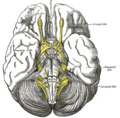 قاعدة المخ.
 -
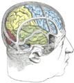 رسم يوضح العلاقة بين المخ والجمجمة.
 -
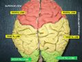
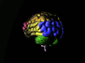 الفص القذالي بالأزرق.
 -
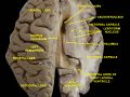
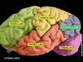 القص القذالي.
 -
 القص القذالي.
 -
 Ventricles of brain and basal ganglia.Superior view. Horizontal section.Deep dissection


انظر أيضاً
هذا المقال يستخدم مصطلحات التشريح؛ لنظرة عامة، انظر مصطلحات التشريح.
- فصوص المخ
- مناطق المخ البشري
- Lunate sulcus
- Visual evoked potential
- Vertical occipital fasciculus
- Visual snow syndrome
المصادر
- ^ "SparkNotes: Brain Anatomy: Parietal and Occipital Lobes". Archived from the original on 2007-12-31. Retrieved 2008-02-27.
- ^ الدماغ، طب تايم
- ^ أ ب ت ث ج ح خ د خطأ استشهاد: وسم
<ref>غير صحيح؛ لا نص تم توفيره للمراجع المسماةrehman - ^ Schacter, D. L., Gilbert, D. L. & Wegner, D. M. (2009). Psychology. (2nd ed.). New York: Worth Publishers.
- ^ الفص الصدغي والفص القفوي، القذالي: الوظائف والتغيرات مع تقدم العمر، طبيب دوت كوم
- ^ Carlson, Neil R. (2007). Psychology : the science of behaviour. New Jersey, USA: Pearson Education. pp. 115. ISBN 978-0-205-64524-4.
- ^ Chilosi, Anna Maria; Brovedani (November 2006). "Neuropsychological Findings in Idiopathic Occipital Lobe Epilepsies". Epilepsia. 47 (s2): 76–78. doi:10.1111/j.1528-1167.2006.00696.x. PMID 17105468. S2CID 23702191.
- ^ Destina Yalçin, A.; Kaymaz, A.; Forta, H. (2000). "Reflex occipital lobe epilepsy". Seizure. 9 (6): 436–441. doi:10.1053/seiz.2000.0424. PMID 10986003.
- ^ أ ب ت Adcock, Jane E; Panayiotopoulos, Chrysostomos P (31 October 2012). "Journal of Clinical Neurophysiology". Occipital Lobe Seizures and Epilepsies. 29 (5): 397–407. doi:10.1097/wnp.0b013e31826c98fe. PMID 23027097.