كراتين

الكـِراتين أو القرتين ( Keratin ؛ /ˈkɛrətɪn/[1][2])، هي عائلة من الپروتينات الليفية، وهي صلبة وعديمة الذوبان موجودة لدى الزواحف والطيور والبرمائيات والثدييات.[3][4] وتدخل بشكل أساسي في تكوين الشعر والصوف والأظافر والمخالب والجلد ومينا الأسنان والحوافر والقرون، لا يضاهيها في الصلابة من بين المواد الحيوية سوى الكايتين Chitin.[5][6][7] ويأتي الكيراتين على نوعين: البدائي، في الصيَغ الأطرى الموجودة في كل الفقاريات، والأصلب، في الصيَغ المشتقة الموجودة فقط في سحليات الوجه (الزواحف والطيور).
تعريف
الكراتين هو پروتين متين عديم الذوبان يوجد في الطبقة الخارجية للجلد عند بني البشر وحيوانات أخرى عديدة. وتدعى هذه الطبقة الخارجية البشرة. تحتوي الطبقة السطحية من خلايا البشرة على القرَتين. ويجعل القرتين الموجود في هذه الخلايا الجلد متينًا ويكاد يكون مانعًا للماء بشكل تام. ويزداد عدد الخلايا المحتوية على القرتين في الأماكن التي يتعرض فيها الجلد لكثير من الاحتكاك والارتخاء ـ مثل الكفين والقدمين ـ مما يؤدي إلى تكون ما يسمى الجسأة. ويتم طرح الخلايا المحتوية على القرتين واستبدال أخرى جديدة بها باستمرار. وتحدث الحالة المعروفة باسم الهبرية، عندما تطرح فروة الرأس مثل هذه الخلايا.
أمثلة التواجد

Alpha-keratins (α-keratins) are found in all vertebrates. They form the hair (including wool), the outer layer of skin, horns, nails, claws and hooves of mammals, and the slime threads of hagfish.[4] The baleen plates of filter-feeding whales are also made of keratin. Keratin filaments are abundant in keratinocytes in the hornified layer of the epidermis; these are proteins which have undergone keratinization. They are also present in epithelial cells in general. For example, mouse thymic epithelial cells react with antibodies for keratin 5, keratin 8, and keratin 14. These antibodies are used as fluorescent markers to distinguish subsets of mouse thymic epithelial cells in genetic studies of the thymus.
The harder beta-keratins (β-keratins) are found only in the sauropsids, i.e., all living reptiles and birds. They are found in the nails, scales, and claws of reptiles, in some reptile shells (Testudines), and in the feathers, beaks, and claws of birds.[8] These keratins are formed primarily in beta sheets. However, beta sheets are also found in α-keratins.[9] Recent scholarship has shown that sauropsid β-keratins are fundamentally different from α-keratins at a genetic and structural level. The new term corneous beta protein (CBP) has been proposed to avoid confusion with α-keratins.[10]
Keratins (also described as cytokeratins) are polymers of type I and type II intermediate filaments that have been found only in chordates (vertebrates, amphioxi, urochordates).[مطلوب توضيح] Nematodes and many other non-chordate animals seem to have only type V intermediate filaments, fibers that structure the nucleus.
الجينات


The human genome encodes 54 functional keratin genes, located in two clusters on chromosomes 12 and 17. This suggests that they originated from a series of gene duplications on these chromosomes.[11]
The keratins include the following proteins of which KRT23, KRT24, KRT25, KRT26, KRT27, KRT28, KRT31, KRT32, KRT33A, KRT33B, KRT34, KRT35, KRT36, KRT37, KRT38, KRT39, KRT40, KRT71, KRT72, KRT73, KRT74, KRT75, KRT76, KRT77, KRT78, KRT79, KRT80, KRT81, KRT82, KRT83, KRT84, KRT85 and KRT86 have been used to describe keratins past 20.[12]
| الرمز | العملية الحيوية |
|---|---|
| KRT1 | complement activation, lectin pathway |
| KRT1 | retina homeostasis |
| KRT1 | response to oxidative stress |
| KRT1 | peptide cross-linking |
| KRT1 | keratinization |
| KRT1 | fibrinolysis |
| KRT1 | intermediate filament organization |
| KRT1 | regulation of angiogenesis |
| KRT1 | negative regulation of inflammatory response |
| KRT1 | protein heterotetramerization |
| KRT1 | establishment of skin barrier |
| KRT10 | morphogenesis of an epithelium |
| KRT10 | epidermis development |
| KRT10 | peptide cross-linking |
| KRT10 | keratinocyte differentiation |
| KRT10 | epithelial cell differentiation |
| KRT10 | positive regulation of epidermis development |
| KRT10 | protein heterotetramerization |
| KRT12 | morphogenesis of an epithelium |
| KRT12 | visual perception |
| KRT12 | epidermis development |
| KRT12 | epithelial cell differentiation |
| KRT12 | cornea development in camera-type eye |
| KRT13 | cytoskeleton organization |
| KRT13 | epithelial cell differentiation |
| KRT13 | regulation of translation in response to stress |
| KRT13 | intermediate filament organization |
| KRT14 | aging |
| KRT14 | epidermis development |
| KRT14 | keratinocyte differentiation |
| KRT14 | epithelial cell differentiation |
| KRT14 | hair cycle |
| KRT14 | intermediate filament organization |
| KRT14 | intermediate filament bundle assembly |
| KRT14 | stem cell differentiation |
| KRT15 | epidermis development |
| KRT15 | epithelial cell differentiation |
| KRT15 | intermediate filament organization |
| KRT16 | morphogenesis of an epithelium |
| KRT16 | inflammatory response |
| KRT16 | cytoskeleton organization |
| KRT16 | aging |
| KRT16 | keratinocyte differentiation |
| KRT16 | negative regulation of cell migration |
| KRT16 | epithelial cell differentiation |
| KRT16 | keratinization |
| KRT16 | hair cycle |
| KRT16 | innate immune response |
| KRT16 | intermediate filament cytoskeleton organization |
| KRT16 | intermediate filament organization |
| KRT16 | keratinocyte migration |
| KRT16 | establishment of skin barrier |
| KRT17 | morphogenesis of an epithelium |
| KRT17 | positive regulation of cell growth |
| KRT17 | epithelial cell differentiation |
| KRT17 | hair follicle morphogenesis |
| KRT17 | keratinization |
| KRT17 | intermediate filament organization |
| KRT17 | positive regulation of translation |
| KRT17 | positive regulation of hair follicle development |
| KRT18 | cell cycle |
| KRT18 | anatomical structure morphogenesis |
| KRT18 | tumor necrosis factor-mediated signaling pathway |
| KRT18 | obsolete Golgi to plasma membrane CFTR protein transport |
| KRT18 | Golgi to plasma membrane protein transport |
| KRT18 | negative regulation of apoptotic process |
| KRT18 | intermediate filament cytoskeleton organization |
| KRT18 | extrinsic apoptotic signaling pathway |
| KRT18 | hepatocyte apoptotic process |
| KRT18 | cell-cell adhesion |
| KRT19 | Notch signaling pathway |
| KRT19 | epithelial cell differentiation |
| KRT19 | response to estrogen |
| KRT19 | intermediate filament organization |
| KRT19 | sarcomere organization |
| KRT19 | cell differentiation involved in embryonic placenta development |
| KRT2 | keratinocyte development |
| KRT2 | epidermis development |
| KRT2 | peptide cross-linking |
| KRT2 | keratinization |
| KRT2 | keratinocyte activation |
| KRT2 | keratinocyte proliferation |
| KRT2 | intermediate filament organization |
| KRT2 | positive regulation of epidermis development |
| KRT2 | keratinocyte migration |
| KRT20 | apoptotic process |
| KRT20 | cellular response to starvation |
| KRT20 | epithelial cell differentiation |
| KRT20 | intermediate filament organization |
| KRT20 | regulation of protein secretion |
| KRT23 | epithelial cell differentiation |
| KRT23 | intermediate filament organization |
| KRT24 | biological_process |
| KRT25 | cytoskeleton organization |
| KRT25 | aging |
| KRT25 | hair follicle morphogenesis |
| KRT25 | hair cycle |
| KRT25 | intermediate filament organization |
| KRT26 | |
| KRT27 | biological_process |
| KRT27 | hair follicle morphogenesis |
| KRT27 | intermediate filament organization |
| KRT28 | biological_process |
| KRT3 | epithelial cell differentiation |
| KRT3 | keratinization |
| KRT3 | intermediate filament cytoskeleton organization |
| KRT3 | intermediate filament organization |
| KRT31 | epidermis development |
| KRT31 | epithelial cell differentiation |
| KRT31 | intermediate filament organization |
| KRT32 | epidermis development |
| KRT32 | epithelial cell differentiation |
| KRT32 | intermediate filament organization |
| KRT33A | epithelial cell differentiation |
| KRT33A | intermediate filament organization |
| KRT33B | aging |
| KRT33B | epithelial cell differentiation |
| KRT33B | hair cycle |
| KRT33B | intermediate filament organization |
| KRT34 | epidermis development |
| KRT34 | epithelial cell differentiation |
| KRT34 | intermediate filament organization |
| KRT35 | anatomical structure morphogenesis |
| KRT35 | epithelial cell differentiation |
| KRT35 | intermediate filament organization |
| KRT36 | biological_process |
| KRT36 | epithelial cell differentiation |
| KRT36 | intermediate filament organization |
| KRT36 | regulation of keratinocyte differentiation |
| KRT37 | epithelial cell differentiation |
| KRT37 | intermediate filament organization |
| KRT38 | epithelial cell differentiation |
| KRT38 | intermediate filament organization |
| KRT39 | epithelial cell differentiation |
| KRT39 | intermediate filament organization |
| KRT4 | cytoskeleton organization |
| KRT4 | epithelial cell differentiation |
| KRT4 | keratinization |
| KRT4 | intermediate filament organization |
| KRT4 | negative regulation of epithelial cell proliferation |
| KRT40 | epithelial cell differentiation |
| KRT40 | intermediate filament organization |
| KRT5 | epidermis development |
| KRT5 | response to mechanical stimulus |
| KRT5 | regulation of cell migration |
| KRT5 | keratinization |
| KRT5 | regulation of protein localization |
| KRT5 | intermediate filament polymerization |
| KRT5 | intermediate filament organization |
| KRT6A | obsolete negative regulation of cytolysis by symbiont of host cells |
| KRT6A | morphogenesis of an epithelium |
| KRT6A | positive regulation of cell population proliferation |
| KRT6A | cell differentiation |
| KRT6A | keratinization |
| KRT6A | wound healing |
| KRT6A | intermediate filament organization |
| KRT6A | defense response to Gram-positive bacterium |
| KRT6A | cytolysis by host of symbiont cells |
| KRT6A | antimicrobial humoral immune response mediated by antimicrobial peptide |
| KRT6A | negative regulation of entry of bacterium into host cell |
| KRT6B | ectoderm development |
| KRT6B | keratinization |
| KRT6B | intermediate filament organization |
| KRT6C | keratinization |
| KRT6C | intermediate filament cytoskeleton organization |
| KRT6C | intermediate filament organization |
| KRT7 | keratinization |
| KRT7 | intermediate filament organization |
| KRT71 | hair follicle morphogenesis |
| KRT71 | keratinization |
| KRT71 | intermediate filament organization |
| KRT72 | biological_process |
| KRT72 | keratinization |
| KRT72 | intermediate filament organization |
| KRT73 | biological_process |
| KRT73 | keratinization |
| KRT73 | intermediate filament organization |
| KRT74 | keratinization |
| KRT74 | intermediate filament cytoskeleton organization |
| KRT74 | intermediate filament organization |
| KRT75 | hematopoietic progenitor cell differentiation |
| KRT75 | keratinization |
| KRT75 | intermediate filament organization |
| KRT76 | cytoskeleton organization |
| KRT76 | epidermis development |
| KRT76 | keratinization |
| KRT76 | pigmentation |
| KRT76 | intermediate filament organization |
| KRT76 | sebaceous gland development |
| KRT77 | biological_process |
| KRT77 | keratinization |
| KRT77 | intermediate filament organization |
| KRT78 | keratinization |
| KRT78 | intermediate filament organization |
| KRT79 | keratinization |
| KRT79 | intermediate filament organization |
| KRT8 | keratinization |
| KRT8 | tumor necrosis factor-mediated signaling pathway |
| KRT8 | intermediate filament organization |
| KRT8 | sarcomere organization |
| KRT8 | response to hydrostatic pressure |
| KRT8 | response to other organism |
| KRT8 | cell differentiation involved in embryonic placenta development |
| KRT8 | extrinsic apoptotic signaling pathway |
| KRT8 | hepatocyte apoptotic process |
| KRT80 | keratinization |
| KRT80 | intermediate filament organization |
| KRT81 | keratinization |
| KRT81 | intermediate filament organization |
| KRT82 | biological_process |
| KRT82 | keratinization |
| KRT82 | intermediate filament organization |
| KRT83 | aging |
| KRT83 | epidermis development |
| KRT83 | keratinization |
| KRT83 | hair cycle |
| KRT83 | intermediate filament organization |
| KRT84 | hair follicle development |
| KRT84 | keratinization |
| KRT84 | nail development |
| KRT84 | intermediate filament organization |
| KRT84 | regulation of keratinocyte differentiation |
| KRT85 | epidermis development |
| KRT85 | keratinization |
| KRT85 | intermediate filament organization |
| KRT86 | keratinization |
| KRT86 | intermediate filament organization |
| KRT9 | spermatogenesis |
| KRT9 | epidermis development |
| KRT9 | epithelial cell differentiation |
| KRT9 | skin development |
| KRT9 | intermediate filament organization |

الوظيفة
Keratin filaments are abundant in keratinocytes in the cornified layer of the epidermis; these are cells which have undergone keratinization. In addition, keratin filaments are present in epithelial cells in general. For example, mouse thymic epithelial cells (TECs) are known to react with antibodies for keratin 5, keratin 8, and keratin 14. These antibodies are used as fluorescent markers to distinguish subsets of TECs in genetic studies of the thymus.
- the α-keratins in the hair (including wool), horns, nails, claws and hooves of mammals[التحقق مطلوب]
- the harder β-keratins found in nails and in the scales and claws of reptiles, their shells (chelonians, such as tortoise, turtle, terrapin), and in the feathers, beaks, claws of birds and quills of porcupines.[14] (These keratins are formed primarily in beta sheets. However, beta sheets are also found in α-keratins.)[15]
The baleen plates of filter-feeding whales are made of keratin.
Although it is now difficult to be certain, the scales, claws, some protective armour and the beaks of dinosaurs were probably composed of keratin.[16]
Keratins (also described as cytokeratins) are polymers of type I and type II intermediate filaments, which have only been found in the genomes of chordates (vertebrates, Amphioxus, urochordates). Nematodes and many other non-chordate animals seem to only have type V intermediate filaments, lamins, which have a long rod domain (vs. a short rod domain for the keratins).
العناية بالشعر
عند فقدان الشعر لمادة الكراتين يبدأ في الجفاف والتقصف وتقل نسبة الكيراتين تدريجيا حتى تصل نسبته في الشعر إلى 35% وهذا لعدة أسباب فأما لأسباب وراثية أو نتيجة فرد الشعر وكثرة الصبغات واستخدام الكريمات والشامبوهات الغلط والاكثار من السيشوار والمكواة ولهذا كان أستخدام الكيراتين المصنع من مواد طبيعية ليقوي نسبة الكيراتين الطبيعية الموجودة في الشعر وبالتالى يرجع الشعر إلى طبيعته وصحته فمادة الكيراتين هي المسئولة عن صلابة الشعر ومقاومته للحرارة اللي ممكن يتعرض لها من العوامل الخارجية نتيجة كثرة استعمال المكواة والسشوار أو الصبغة الكيماوية ولذلك يعتبر الكيراتين فرد وعلاج للشعر وهو أحدث علاج للشعر التالف المقصف أو المجعد أو الناشف أو المتساقط أو الشعر المجهد ويتغلغل في الأماكن التالفة ويعالجها وميزاته إنه غير قابل للذوبان ويصلح لجميع أنواع الشعر وكل الأعمار.[17]
الأنواع المستخدمة
الكيراتين المستخدم للعناية بالشعر، له أنواع من حيث مدة مفعوله ومن حيث الغرض منه فيل أنه 7 أنواع كل نوع بيناسب طبيعة كل شعر فيوجد كيراتين فرد وعلاج وهو أغلى الأنواع ويوجد كيراتين لفرد فقط أو مايسمى الليس وهناك نوع لعلاج فقط أو ما يسمى تريتمنت ويوجد نوع يستخدم مرة واحدة وتستمر نتائجة للأبد لأنه يرفع نسبة الكيراتين الطبيعية في الجسم ويوجد نوع مؤقت اى يدوم فترة ثم يحتاج التكرار كل فترة ونتيجته بتستمر من 3 شهور إلى 3 سنين وعموما درجة الفرد والتموج حسب الرغبة فأما لفرد الخالص أو الفرد بدرجه بها تموجات واسعة ويوجد الآن بدائل لكيراتين وكلها من منتجات الكيراتين المعالج أو الترتمنت وهى أرخص نسبيا عن الكيراتين ككريم كيراتينو وكريم Rb Hot Couffier.
مادة الفورمالين
الكيراتين المقلد يحتوى على مادة الفورمالين وهى مادة مسرطنة ويتم إضافتها بنسبة كبيرة جدا ويضاف الكثير من المواد الكيماوية والبطاس وهى مواد خطيرة جدا على الشعر ورائحته مثيرة للدموع وتسبب الشعور بحرقان بسبب المواد الكيماوية الشديدة به ويستمر على الشعر 3 شهور فقط والنوع المقلد بيطلب تتجنب نزول حمام السباحة وثمنه 1500 جنيه مصري أو أقل. أما الآصلى ليس له أي آثار جانبية فملمسه واحد ومتجانس ومن غير حبيبات وخالى من الفورمالين وحاصل على علامة موافقة منظمة الدواء والغذاء الأمريكية.
طريقة الاستخدام
يتم دخول الكيراتين إلى الشعر عن طريق جهاز معين بواسطة أمبولات ويخرج الكيراتين على شكل بخار ويتم تصفيف الشعر به.
البيولوجيا الجزيئية والكيمياء الحيوية
The usefulness of keratins depends on their supermolecular aggregation. These depend on the properties of the individual polypeptide strands, which depend in turn on their amino acid composition and sequence. The α-helix and β-sheet motifs, and disulfide bridges, are crucial to the conformations of globular, functional proteins like enzymes, many of which operate semi-independently, but they take on a completely dominant role in the architecture and aggregation of keratins.
The alpha keratin helix is not a true alpha helix, as it only has 3.5 residues/turn, where the normal alpha helix has 3.6 residues/turn. This is important for the different helices to form tight disulfide bonds. Also, roughly every seventh residue is a leucine, so they can line up and help the strands stick together through hydrophobic interactions.
التقرن
Cornification is the process of forming an epidermal barrier in stratified squamous epithelial tissue. At the cellular level, cornification is characterised by:
- production of keratin
- production of small proline-rich (SPRR) proteins and transglutaminase which eventually form a cornified cell envelope beneath the plasma membrane
- terminal differentiation
- loss of nuclei and organelles, in the final stages of cornification metabolism ceases and the cells are almost completely filled by keratin
During the process of epithelial التمايز, cells become cornified as keratin protein is incorporated into longer keratin intermediate filaments. Eventually the nucleus and cytoplasmic organelles disappear, metabolism ceases and cells undergo a programmed death as they become fully keratinized. In many other cell types, such as cells of the dermis, keratin filaments and other intermediate filaments function as part of the cytoskeleton to mechanically stabilize the cell against physical stress. It does this through connections to desmosomes, cell-cell junctional plaques, and hemidesmosomes, cell-basement membrane adhesive structures.
Cells in the epidermis contain a structural matrix of keratin, which makes this outermost layer of the skin almost waterproof, and along with collagen and elastin, gives skin its strength. Rubbing and pressure cause thickening of the outer, cornified layer of the epidermis and form protective calluses — useful for athletes and on the fingertips of musicians who play stringed instruments. Keratinized epidermal cells are constantly shed and replaced (see dandruff).
These hard, integumentary structures are formed by intercellular cementing of fibers formed from the dead, cornified cells generated by specialized beds deep within the skin. Hair grows continuously and feathers moult and regenerate. The constituent proteins may be phylogenetically homologous but differ somewhat in chemical structure and supermolecular organization. The evolutionary relationships are complex and only partially known. Multiple genes have been identified for the β-keratins in feathers, and this is probably characteristic of all keratins.
التفاصيل البنائية

Fibrous keratin molecules supercoil to form a very stable, left-handed superhelical motif to multimerise, forming filaments consisting of multiple copies of the keratin monomer.[18]
Limited interior space is the reason why the triple helix of the (unrelated) structural protein collagen, found in skin, cartilage and bone, likewise has a high percentage of glycine. The connective tissue protein elastin also has a high percentage of both glycine and alanine. Silk fibroin, considered a β-keratin, can have these two as 75–80% of the total, with 10–15% serine, with the rest having bulky side groups. The chains are antiparallel, with an alternating C → N orientation.[19] A preponderance of amino acids with small, nonreactive side groups is characteristic for structural proteins, for which H-bonded close packing is more important than chemical specificity.
جسور الثاني كبريتيد
In addition to intra- and intermolecular hydrogen bonds, the distinguishing feature of keratins is the presence of large amounts of the sulfur-containing amino acid cysteine, required for the disulfide bridges that confer additional strength and rigidity by permanent, thermally stable crosslinking[20]—in much the same way that non-protein sulfur bridges stabilize vulcanized rubber. Human hair is approximately 14% cysteine. The pungent smells of burning hair and skin are due to the volatile sulfur compounds formed. Extensive disulfide bonding contributes to the insolubility of keratins, except in a small number of solvents such as dissociating or reducing agents.

The more flexible and elastic keratins of hair have fewer interchain disulfide bridges than the keratins in mammalian fingernails, hooves and claws (homologous structures), which are harder and more like their analogs in other vertebrate classes.[21] Hair and other α-keratins consist of α-helically coiled single protein strands (with regular intra-chain H-bonding), which are then further twisted into superhelical ropes that may be further coiled. The β-keratins of reptiles and birds have β-pleated sheets twisted together, then stabilized and hardened by disulfide bridges.
Thiolated polymers (thiomers) can form disulfide bridges with cysteine substructures of keratins getting covalently attached to these proteins.[22] Thiomers therefore exhibit high binding properties to keratins found in hair,[23] on skin[24][25] and on the surface of many cell types.[26]
تشكل الخيوط
It was theorized that keratins are combined into 'hard' and 'soft,' or 'cytokeratins' and 'other keratins'[مطلوب توضيح]. That model is now understood to be correct. A new nuclear addition in 2006 to describe keratins takes this into account.[27]
Keratin filaments are intermediate filaments. Like all intermediate filaments, keratin proteins form filamentous polymers in a series of assembly steps beginning with dimerization; dimers assemble into tetramers and octamers and eventually, the current hypothesis holds, into unit-length-filaments (ULF) capable of annealing end-to-end into long filaments.
الاقتران
| أ (طبيعي-قاعدي) | ب (حامضي) | التواجد |
|---|---|---|
| كراتين 1، كراتين 2 | كراتين 9، كراتين 10 | الطبقة القرنية، الخلايا الكراتينية |
| كراتين 3 | كراتين 12 | القرنية |
| كراتين 4 | كراتين 13 | ظهارة مطبقة الخلايا |
| كراتين 5 | كراتين 14، كراتين 15 | ظهارة مطبقة الخلايا |
| كراتين 6 | كراتين 16، كراتين 17 | ظهارة حرشفية الخلايا |
| كراتين 7 | كراتين 19 | ductal epithelia |
| كراتين 8 | كراتين 18، كراتين 20 | ظهارة بسيطة |
الحرير
fibroins الحرير التي تنتجها الحشرات والعناكب كثراً ما تـُصنف ككراتينات، بالرغم من أنه غير واضح إذا ما كانت phylogenetically related لكراتينات الفقاريات.
Silk found in insect pupae, and in spider webs and egg casings, also has twisted β-pleated sheets incorporated into fibers wound into larger supermolecular aggregates. The structure of the spinnerets on spiders’ tails, and the contributions of their interior glands, provide remarkable control of fast extrusion. حرير العنكبوت عادة ما يكون سمكه 1 - 2 ميكرومتر (µm)، بالمقارنة بـ 60 µm للشعر البشري، وأكثر لبعض الثدييات. السمات المفيدة حيوياً وتجارياً لألياف الحرير تعتمد على تنظيم يلاسل بروتين متجاورة متعددة في مناطق بلورية صلبة بأحجام متغيرة متبادلة مع مناطق غير متبلورة مرنة حيث تكون السلاسل randomly coiled.[28] A somewhat analogous situation occurs with synthetic بوليمرات مثل النايلون، developed as a silk substitute. الحرير من hornet cocoon contains doublets about 10 µm across, with cores and coating, and may be arranged in up to 10 layers; also in plaques of variable shape. Adult hornets also use silk as a glue, as do spiders.
الأهمية السريرية
بعض الفطريات المعدية، مثل أولئك المتسببين في قدم الرياضي، الدودة الحلقية، or the Batrachochytrium dendrobatidis (Chytrid fungus) which is killing amphibians all over the world, feed on keratin.
الأمراض التي تسببها التحورات في جينات الكراتين تضم
- Epidermolysis bullosa simplex
- Ichthyosis bullosa of Siemens
- Epidermolytic hyperkeratosis
- Steatocystoma multiplex
- Keratosis pharyngis
أنظر أيضاً
صور اضافية
-
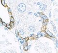 Keratin (high molecular weight) in bile duct cell and oval cells of mouse liver.
المصادر
- ^ OED 2nd edition, 1989 as /ˈkɛrətɪn/
- ^ Entry "keratin" Archived 2013-05-09 at the Wayback Machine in Merriam-Webster Online Dictionary Archived 2017-09-22 at the Wayback Machine.
- ^ Fraser R (1972). Keratins: Their composition, structure and biosynthesis. Bannerstone House: Charles C Thomas. pp. 3–6. ISBN 978-0-398-02283-9.
- ^ أ ب Wang B, Yang W, McKittrick J, Meyers MA (March 2016). "Keratin: Structure, mechanical properties, occurrence in biological organisms, and efforts at bioinspiration". Progress in Materials Science. 76: 229–318. doi:10.1016/j.pmatsci.2015.06.001.
- ^ "Keratin". Webster's Online Dictionary. 22 May 2023. Archived from the original on 1 May 2021. Retrieved 9 August 2018.
- ^ Vincent JF, Wegst UG (July 2004). "Design and mechanical properties of insect cuticle". Arthropod Structure & Development. 33 (3): 187–199. Bibcode:2004ArtSD..33..187V. doi:10.1016/j.asd.2004.05.006. PMID 18089034.
- ^ Tombolato L, Novitskaya EE, Chen PY, Sheppard FA, McKittrick J (February 2010). "Microstructure, elastic properties and deformation mechanisms of horn keratin". Acta Biomaterialia. 6 (2): 319–330. doi:10.1016/j.actbio.2009.06.033. PMID 19577667.
- ^ Hickman CP, Roberts LS, Larson AL (2003). Integrated principles of zoology. Dubuque, IA: McGraw-Hill. pp. 538. ISBN 978-0-07-243940-3.
- ^ Kreplak L, Doucet J, Dumas P, Briki F (July 2004). "New Aspects of the α-Helix to β-Sheet Transition in Stretched Hard α-Keratin Fibers". Biophysical Journal. 87 (1): 640–647. Bibcode:2004BpJ....87..640K. doi:10.1529/biophysj.103.036749. PMC 1304386. PMID 15240497.
- ^ Alibardi L (September 2016). "Sauropsids Cornification is Based on Corneous Beta-Proteins, a Special Type of Keratin-Associated Corneous Proteins of the Epidermis". Journal of Experimental Zoology. Part B, Molecular and Developmental Evolution. 326 (6): 338–351. Bibcode:2016JEZB..326..338A. doi:10.1002/jez.b.22689. PMID 27506161.
- ^ Moll R, Divo M, Langbein L (June 2008). "The human keratins: biology and pathology". Histochemistry and Cell Biology. 129 (6) 705: 705–733. doi:10.1007/s00418-008-0435-6. PMC 2386534. PMID 18461349.
- ^ Schweizer J, Bowden PE, Coulombe PA, Langbein L, Lane EB, Magin TM, et al. (July 2006). "New consensus nomenclature for mammalian keratins". The Journal of Cell Biology. 174 (2): 169–174. doi:10.1083/jcb.200603161. PMC 2064177. PMID 16831889.
- ^ "GeneCards - Human Genes | Gene Database". Archived from the original on 2023-05-13. Retrieved 2023-05-08.
- ^ Hickman, Cleveland Pendleton; Roberts, Larry S.; Larson, Allan L. (2003). Integrated principles of zoology. Dubuque, IA: McGraw-Hill. p. 538. ISBN 0-07-243940-8.
{{cite book}}: Cite has empty unknown parameter:|coauthors=(help)CS1 maint: multiple names: authors list (link) - ^ Kreplak L, Doucet J, Dumas P, Briki F (2004). "New aspects of the alpha-helix to beta-sheet transition in stretched hard alpha-keratin fibers". Biophys J. 87 (1): 640–7. doi:10.1529/biophysj.103.036749. PMC 1304386. PMID 15240497.
{{cite journal}}: CS1 maint: multiple names: authors list (link) - ^ http://www.ncbi.nlm.nih.gov/pmc/articles/PMC1617199/
- ^ الكيراتين وأنواعه وأشهر الأسئلة عنه، موسوعة الجمال
- ^ {{{author}}}, Fundamentals of Biochemistry, [[{{{publisher}}}]], [[{{{date}}}]].
- ^ "Secondary Protein". Elmhurst.edu. Retrieved 2010-09-23.
- ^ "What is Keratin?". WiseGEEK. Archived from the original on 13 May 2014. Retrieved 11 May 2014.
- ^ H Bragulla H, G Homberger D (Apr 2009). "Structure and functions of keratin proteins in simple, stratified, keratinized and cornified epithelia". Journal of Anatomy. 214 (4): 516–559. doi:10.1111/j.1469-7580.2009.01066.x. PMC 2736122. PMID 19422428.
- ^ Leichner C, Jelkmann M, Bernkop-Schnürch A (2019). "Thiolated polymers: Bioinspired polymers utilizing one of the most important bridging structures in nature". Advanced Drug Delivery Reviews. 151–152: 191–221. doi:10.1016/j.addr.2019.04.007. PMID 31028759.
- ^ {{{1}}} patent {{{2}}}
- ^ Grießinger J, Bonengel S, Partenhauser A, Ijaz M, Bernkop-Schnürch A (2017). "Thiolated polymers: Evaluation of their potential as dermoadhesive excipients". Drug Development and Industrial Pharmacy. 43 (2): 204–212. doi:10.1080/03639045.2016.1231809. PMID 27585266.
- ^ Partenhauser A, Zupančič O, Rohrer J, Bonengel S, Bernkop-Schnürch A (2015). "Thiolated silicone oils as adhesive skin protectants for improved barrier function". International Journal of Cosmetic Science. 38 (3): 257–265. doi:10.1111/ics.12284. PMID 26444859.
- ^ Le-Vinh B, Steinbring C, Nguyen Le N, Matuszczak B, Bernkop-Schnürch A (2023). "S-Protected thiolated chitosan versus thiolated chitosan as cell adhesive biomaterials for tissue engineering". ACS Applied Materials & Interfaces. 15 (34): 40304–40316. doi:10.1021/acsami.3c09337. PMC 10472333. PMID 37594415.
- ^ Schweizer J, Bowden PE, Coulombe PA, Langbein L, Lane EB, Magin TM, Maltais L, Omary MB, Parry DA, Rogers MA, Wright MW. New consensus nomenclature for mammalian keratins. J Cell Biol. 2006 Jul 17;174(2):169-74.
- ^ Australia. "Spiders - Silk structure". Amonline.net.au. Retrieved 2010-09-23.
للاستزادة
Ramot Y, Paus R, Tiede S, Zlotogorski A. Endocrine controls of keratin expression. Bioessays. 2009 Apr;31(4):389-99. PMID: 19274655
وصلات خارجية
- Pages with plain IPA
- Short description is different from Wikidata
- Articles with hatnote templates targeting a nonexistent page
- جميع الصفحات التي تحتاج تنظيف
- مقالات بالمعرفة تحتاج توضيح from June 2026
- مقالات بالمعرفة تحتاج تمحيص الحقائق from November 2007
- جميع الصفحات التي تحتاج تمحيص حقائق
- مقالات بالمعرفة تحتاج توضيح from May 2010
- كراتينات
- هيكل خلوي