كتف
| الكتف Shoulder | |
|---|---|
 | |
 كبسولة مفصل الكتف. صورة أمامية. | |
| Details | |
| المُعرفات | |
| اللاتينية | articulatio humeri |
| MeSH | D012782 |
| TA98 | A01.1.00.020 |
| TA2 | 139 |
| FMA | 25202 |
| المصطلحات التشريحية | |
الكتف إنگليزية: Shoulder هي المنطقة التي تربط الذراع مع الجذع. تتكون من 3 عظام الترقوة ولوح الكتف والجزء العلوي من عظمة العضد.
يحيط بالمفصل الكبسولة. وتحيط اربطة بالكبسولة، والاربطة تكون خفيفة وترتبط قريبا جدا من سطح التمفصل، عدا في الأسفل فإنها تمتد لتلتصق بالرقبة الجراحية لعظم العضد (عنق العضد الجراحي) لذلك تسمح هذه الأربطة في أسفل مفصل الكتف بالحركة الواسعة برفع الذراع إلى الأعلى.
أربطة الكبسولة : هناك ثلاثة أربطة ليست قوية جداً من الأمام الأربطة شبه الحقية العضدية. وهناك رباط من فوق وفيه فتحتين مما يضعفه أكثر الرباط الغرابي العضدي.
أمام المفصل هناك وتر ما تحت الكبسولة، وبينه وبين المفصل كيس صغير هو كيس المفصل تحت الكبسولة، وهذا لديه اتصال مع داخل المفصل.
التركيب
مفصل الكتف

مفصل الكتف هو مفصل في الجسم من نوع للمفصل كرة وياالحُقّي تكون الكرة هنا كبيرة الحجم والحفرة ضحلة. و التالي فانه يكون معرُض ّللخلع (أي خروج رأس العظم من الحفرة التي يجلس فيها). وكما هو الحال في بقية المفاصل من نفس النوع فان المناطق المتلامسة في مفصل الكتف تكون مغطاة بغضروف من النوع الهلامي لكي تسهل الحركة بين العظام المتلامسة في المفصل.
الكفة المدورة


الكفة المدورة هي مصطلح تشريحي يشمل مجموعة من العضلات والأوتار الخاصة بها التي تقوم بتثبيت الكتف.[1][2][3] تشكل عضلات الكفة المدورة الأربعة مع العضلتين الدالية والمدورة الكبيرة ما يسمى بالعضلات الكتفية العضدية.
عضلات أخرى
| الاسم | الارتباط | الوظيفة |
| serratus anterior | Originates on the surface of the upper eight ribs at the side of the chest and inserts along the entire anterior length of the medial border of the scapula.[4] | It fixes the scapula into the thoracic wall and aids in rotation and abduction of the shoulders.[citation needed] |
| subclavius | Located beneath the clavicle, originating from the first rib and inserting on the subclavian groove of the clavicle.[4] | It depresses the lateral clavicle[4] and also acts to stabilize the clavicle.[citation needed] |
| pectoralis minor | Arises from the third, fourth, and fifth ribs, near their cartilage and inserts into the medial border and upper surface of the coracoid process of the scapula.[4] | This muscle aids in respiration, medially rotates the scapula, protracts the scapula, and also draws the scapula inferiorly. |
| sternocleidomastoid | Attaches to the sternum (sterno-), the clavicle (cleido-), and the mastoid process of the temporal bone of the skull. | Most of its actions flex and rotate the head. In regards to the shoulder, however, it also aids in respiration by elevating the sternoclavicular joint when the head is fixed.[citation needed] |
| levator scapulae | Arises from the transverse processes of the first four cervical vertebrae and inserts into the medial border of the scapula. | It is capable of rotating the scapula downward and elevating the scapula.[citation needed] |
العضلات من الظهر
| rhomboid major and rhomboid minor (work together) | They arise from the spinous processes of the thoracic vertebrae T1 to T5 as well as from the spinous processes of the seventh cervical. They attach to the inner border of the scapula.[4] | They are responsible for downward rotation of the scapula with the levator scapulae, as well as adduction of the scapula. |
| trapezius | Arises from the occipital bone, the ligamentum nuchae, the spinous process of the seventh cervical, and the spinous processes of all the thoracic vertebrae.[4] It attaches to the outer clavicle, the acromion process, and into the spine of the scapula.[4] | Different portions of the fibers perform different actions on the scapula: depression, upward rotation, elevation, and retraction.[4] |
| Levator scapula | Arises from the transverse processes of cervical vertebrae 1-4, and attaches to the upper part of the inner border of the scapula.[4] | Elevates the scapula.[4] |
| Latissimus dorsi | A large muscle that arises form the spinous processes of the lower six thoracic vertebrae, lumbar and all sacral vertebrae, and posterior iliac crest. It attaches to the intertubercular groove of the humerus.[4] | Adducts, extends and rotates the humerus inwards.[4] |
الإبط
الإمدادات والممرات العصبية


الأوعية الدموية
_(14764575614).jpg&filetimestamp=20180528015255&)
الوظيفة
| الاسم | الوصف | العضلات |
|---|---|---|
| Scapular retraction [5] (aka scapular adduction) | The scapula is moved posteriorly and medially along the back, moving the arm and shoulder joint posteriorly. Retracting both scapulae gives a sensation of "squeezing the shoulder blades together." | rhomboideus major, minor, and trapezius |
| Scapular protraction[5] (aka scapular abduction) | The opposite motion of scapular retraction. The scapula is moved anteriorly and laterally along the back, moving the arm and shoulder joint anteriorly. If both scapulae are protracted, the scapulae are separated and the pectoralis major muscles are squeezed together.[6] | serratus anterior (prime mover), pectoralis minor and major |
| Scapular elevation [7] | The scapula is raised in a shrugging motion. | levator scapulae, the upper fibers of the trapezius |
| Scapular depression [7] | The scapula is lowered from elevation. The scapulae may be depressed so that the angle formed by the neck and shoulders is obtuse, giving the appearance of "slumped" shoulders.[citation needed] | pectoralis minor, lower fibers of the trapezius, subclavius, latissimus dorsi |
| Arm abduction [8] | Arm abduction occurs when the arms are held at the sides, parallel to the length of the torso, and are then raised in the plane of the torso. This movement may be broken down into two parts: True abduction of the arm, which takes the humerus from parallel to the spine to perpendicular; and upward rotation of the scapula, which raises the humerus above the shoulders until it points straight upwards.[citation needed] | True abduction: supraspinatus (first 15 degrees), deltoid; Upward rotation: trapezius, serratus anterior |
| Arm adduction [9] | Arm adduction is the opposite motion of arm abduction. It can be broken down into two parts: downward rotation of the scapula and true adduction of the arm. | Downward rotation: pectoralis minor, pectoralis major, subclavius, latissimus dorsi (same as scapular depression, with pec major replacing lower fibers of trapezius); True Adduction: latissimus dorsi, subscapularis, teres major, infraspinatus, teres minor, pectoralis major, long head of triceps, coracobrachialis. |
| Arm flexion [10] | The humerus is rotated out of the plane of the torso so that it points forward (anteriorly). | pectoralis major, coracobrachialis, biceps brachii, anterior fibers of deltoid. |
| Arm extension [10] | The humerus is rotated out of the plane of the torso so that it points backwards (posteriorly) | latissimus dorsi and teres major, long head of triceps, posterior fibers of the deltoid |
| Medial rotation of the arm [11] | Medial rotation of the arm is most easily observed when the elbow is held at a 90-degree angle and the fingers are extended so they are parallel to the ground. Medial rotation occurs when the arm is rotated at the shoulder so that the fingers change from pointing straight forward to pointing across the body. | subscapularis, latissimus dorsi, teres major, pectoralis major, anterior fibers of deltoid |
| Lateral rotation of the arm[11] | The opposite of medial rotation of the arm. | infraspinatus and teres minor, posterior fibers of deltoid |
| Arm circumduction[12] | Movement of the shoulder in a circular motion so that if the elbow and fingers are fully extended the subject draws a circle in the air lateral to the body. In circumduction, the arm is not lifted above parallel to the ground so that "circle" that is drawn is flattened on top. | pectoralis major, subscapularis, coracobrachialis, biceps brachii, supraspinatus, deltoid, latissimus dorsi, teres major and minor, infraspinatus, long head of triceps |
الأهمية السريرية

الكسر
الألم
تصوير الكتف
أشعة إكس
-
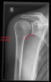 CR. shoulay film.
 -
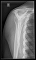
 Transaxillary conventional radiography
 -
 Y-projection conventional radiography


الموجات فوق الصوتية
 |
 |
التصوير بالرنين المغناطيسي
في الحيوانات
معرض الصور
-
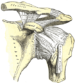 The left shoulder and acromioclavicular joints, and the proper ligaments of the scapula
 -
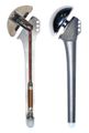 Instrumented shoulder endoprosthesis, with a 9-channel telemetry transmitter to measure six load components in vivo


انظر أيضاً
هذا المقال يستخدم مصطلحات التشريح؛ لنظرة عامة، انظر مصطلحات التشريح.
- Shoulder girdle (Pectoral girdle)
- Sternoclavicular joint
- Chip on shoulder
- Ambe
- Milwaukee shoulder syndrome
المصادر
- ^ Hegedus EJ, Goode A, Campbell S, et al. (February 2008). "Physical examination tests of the shoulder: a systematic review with meta-analysis of individual tests". British Journal of Sports Medicine. 42 (2): 80–92. doi:10.1136/bjsm.2007.038406. PMID 17720798.
- ^ Escamilla RF, Yamashiro K, Paulos L, Andrews JR (2009). "Shoulder muscle activity and function in common shoulder rehabilitation exercises". Sports Med. 39 (8): 663–85. doi:10.2165/00007256-200939080-00004. PMID 19769415.
- ^ Matava MJ, Purcell DB, Rudzki JR (2005). "Partial-thickness rotator cuff tears". Am J Sports Med. 33 (9): 1405–17. doi:10.1177/0363546505280213. PMID 16127127.
- ^ أ ب ت ث ج ح خ د ذ ر ز س خطأ استشهاد: وسم
<ref>غير صحيح؛ لا نص تم توفيره للمراجع المسماة:4 - ^ أ ب "Scapular Protraction and Retraction". University of Michigan Medical School. 2002. Retrieved December 2010.
{{cite web}}: Check date values in:|accessdate=(help) - ^ Modric, Jan. "Shoulder Muscles Functions". ehealthstar.com. Retrieved 11 April 2017.
- ^ أ ب "Scapular Elevation and Depression". University of Michigan Medical School. 2002. Retrieved December 2010.
{{cite web}}: Check date values in:|accessdate=(help) - ^ "Arm Abduction". University of Michigan Medical School. 2002. Retrieved December 2010.
{{cite web}}: Check date values in:|accessdate=(help) - ^ "Arm Adduction". University of Michigan Medical School. 2002. Retrieved December 2010.
{{cite web}}: Check date values in:|accessdate=(help) - ^ أ ب "Arm Flexion and Extension". University of Michigan Medical School. 2002. Retrieved December 2010.
{{cite web}}: Check date values in:|accessdate=(help) - ^ أ ب "Arm Medial and Lateral Rotation". University of Michigan Medical School. 2002. Retrieved December 2010.
{{cite web}}: Check date values in:|accessdate=(help) - ^ "Arm Circumduction". University of Michigan Medical School. 2002. Retrieved December 2010.
{{cite web}}: Check date values in:|accessdate=(help)
وصلات خارجية

- Video of the shoulder carriage in motion
- NIH (article includes text from this source)
- University of Michigan Medical School module on movements of the shoulder, arm, forearm, and hand
- The Solution for Building Bigger Shoulders
الرأس: جمجمة - جبهة – عين – أذن – أنف – فم – لسان – أسنان – فك – فك سفلي – وجه – خد – ذقن

الرقبة: حلق - حنجرة – تفاحة آدم
جذع: كتف – عمود فقري – ثدي – حلمة – صدر – قفص صدري – بطن – سرة
أطراف: ذراع – مرفق – ساعد – رسغ – يد – اصبع (إبهام - سبابة - وسطى - بنصر - خنصر) – رجل – حـِجر Lap – فخذ – ركبة – رَبـْلة الساق Calf muscle – كعب – كاحل – قدم – اصبع قدم (إبهام القدم Hallux)