القولون الصاعد
| Ascending colon | |
|---|---|
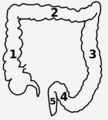
 Drawing of colon seen from front (ascending colon coloured blue) | |
 | |
| Details | |
| السلف | Midgut |
| الشريان | Right colic artery |
| الوريد | Right colic vein |
| العصب | Celiac ganglia, vagus[1] |
| المُعرفات | |
| اللاتينية | colon ascendens |
| MeSH | D044682 |
| TA98 | A05.7.03.002 |
| TA2 | 2982 |
| FMA | 14545 |
| المصطلحات التشريحية | |
In the anatomy of humans and homologous primates, the ascending colon is the part of the colon located between the cecum and the transverse colon.
السمات والبنية
The ascending colon is smaller in calibre than the cecum from where it starts. It passes upward, opposite the colic valve, to the under surface of the right lobe of the liver, on the right of the gall-bladder, where it is lodged in a shallow depression, the colic impression; here it bends abruptly forward and to the left, forming the right colic flexure (hepatic) where it becomes the transverse colon.
It is retained in contact with the posterior wall of the abdomen by the peritoneum, which covers its anterior surface and sides, its posterior surface being connected by loose areolar tissue with the iliacus, quadratus lumborum, aponeurotic origin of transversus abdominis, and with the front of the lower and lateral part of the right kidney.
Sometimes the peritoneum completely invests it and forms a distinct but narrow mesocolon.
It is in relation, in front, with the convolutions of the ileum and the abdominal walls.
Parasympathetic innervation to the ascending colon is supplied by the vagus nerve. Sympathetic innervation is supplied by the thoracic splanchnic nerves.
الموقع
The ascending colon is on the right side of the body (barring any malformations). The term right colon is hypernymous to ascending colon in precise use; many casual mentions of the right colon chiefly concern the ascending colon.

صور إضافية
-
![Inner diameters of different sections of the large intestine, with ascending colon (at left) measuring on average 6.6 cm (range 6.0-7.0 cm).[2]](/w/images/thumb/2/22/Diameters_of_the_large_intestine.svg/120px-Diameters_of_the_large_intestine.svg.png) Inner diameters of different sections of the large intestine, with ascending colon (at left) measuring on average 6.6 cm (range 6.0-7.0 cm).[2]
Inner diameters of different sections of the large intestine, with ascending colon (at left) measuring on average 6.6 cm (range 6.0-7.0 cm).[2] -
-
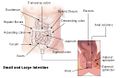 Intestines
 -
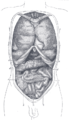 Front view of the thoracic and abdominal viscera.
 -
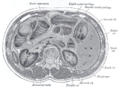
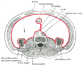 Horizontal disposition of the peritoneum in the lower part of the abdomen.
 -
 The duodenum and pancreas.
 -
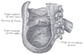 Interior of the cecum and the lower end of ascending colon, showing colic valve.
 -
 Transverse section through the middle of the first lumbar vertebra, showing the relations of the pancreas.
 -
 The relations of the kidneys from behind.
 -
-
 Ascending colon
 -
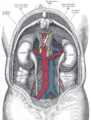
 Mesenteric relation of intestines. Deep dissection. Anterior view.
![Inner diameters of different sections of the large intestine, with ascending colon (at left) measuring on average 6.6 cm (range 6.0-7.0 cm).[2]](/w/index.php?title=%D9%85%D9%84%D9%81:Diameters_of_the_large_intestine.svg&filetimestamp=20230620090210&)


انظر أيضاً
المراجع
'هذه المقالة تَعتمد على مَواد وَمعلومات ذات ملكية عامة، من للطبعة العِشرين لكتاب تشريح گرايز لعام 1918. .
- ^ Nosek, Thomas M. "Section 6/6ch2/s6ch2_30". Essentials of Human Physiology. Archived from the original on 2016-03-24.
- ^ Nguyen H, Loustaunau C, Facista A, Ramsey L, Hassounah N, Taylor H, Krouse R, Payne CM, Tsikitis VL, Goldschmid S, Banerjee B, Perini RF, Bernstein C (2010). "Deficient Pms2, ERCC1, Ku86, CcOI in field defects during progression to colon cancer". J Vis Exp (41). doi:10.3791/1931. PMC 3149991. PMID 20689513.
وصلات خارجية
- SUNY Figs 37:06-08 - "The large intestine."
- Norman/Georgetown largeintestine (cecuminside)