جزر لانگرهانز
| ' | |
|---|---|
 جزر لانگرهانس في فأر، in its typical proximity to a blood vessel; الإنسولين باللون الأحمر، النوى باللون الأزرق. | |
 | |
| Details | |
| Identifiers | |
| اللاتينية | insulae pancreaticae |
| MeSH | D007515 |
| TA98 | A05.9.01.019 |
| TA2 | 3128 |
| FMA | 16016 |
| المصطلحات التشريحية | |


جزر لانگرهانس islets of Langerhans، مناطق في الپنكرياس تحتوي على غدد صماء وظيفتها إفراز الهرمونات. اكتشفها عالم التشريح المرضي الألماني پول لانگرهانس في عام 1869 حيث كان في الثانية والعشرين من عمره،[1] وتشكل جزر لانگرهانس حوالي من 1 إلى 2% من كتلة الپنكرياس. وهذه الجزر غير مزودة بأقنية مفرغة بل تلقي مفرزاتها في الأوعية الدموية مباشرة لذا تدعى داخلية الإفراز أو صماء. [2]
وتظهر جزر لانگرهانس على هيئة بقع صغيرة مختلفة في الشكل والوظيفة عما حولها من خلايا الپنكرياس ولذلك تم تسميتها بالجزر.
التشريح
أنواع خلايا الجزر
- خلايا ألفا وتنتج الگلوكاگون (وتشكل 15–20% من إجمالي عدد خلايا الجزر)، ووظيفة الگلوكاگون اطلاق سكر الگلوكوز المخزن في الكبد والعضلات ليرتفع منسوب السكر في الدم ويغطي حاجة الجسم من الجلوكوز ويتم إفراز الجلوكاجون عادة في أوقات الجوع حيث تنخفض نسبة الجلوكوز الموجودة في الدم عن الحد الأدنى لها. ويعتبر انخفاض منسوب الجلوكوز في الدم في أوقات الجوع هو الحافز الرئيسي لإفراز هرمون الجلوكاجون من خلايا لانگرهانس.
- خلايا بيتا وتنتج الإنسولين والأمايلين (65–80%)، والإنسولين هو الهرمون الذي يفرز عادة بعد تناول الطعام وامتصاص النشويات والسكريات في الجهاز الهضمي ،حيث إن ارتفاع منسوب السكر( الجلوكوز) في الدم بعد تناول الطعام والامتصاص هو العامل الحفاز لإفراز الإنسولين من جزر لانگرهانس. ويعمل هرمون الإنسولين على خفض منسوب السكر في الدم وذلك عن طريق تخزينه في الكبد والعضلات على هيئة النشا الحيواني والمعروف بالجليكوجين.
- خلايا دلتا وتنتج السوماتوستاتين (3–10%)، ويعمل السوماتوستاتين على تقليل النشاط الهضمي في المعدة والأمعاء.
- خلايا پ پ الپپتيد المتعدد الپنكرياسي (3–5%)
- خلايا إپسيلون وتنتج الگريلين (<1%)
تغذية نظير الصماوي
النشاط الكهربي
زرع جزر لانگرهانس كعلاج لمرض السكري النمط الأول
Islet cell transplantation has the possibility of restoring beta cell function from diabetes, offering an alternative to a complete pancreas transplantation or an artificial pancreas.
Because the beta cells in the islets of Langerhans are selectively destroyed by an autoimmune process in type 1 diabetes, clinicians and researchers are actively pursuing islet transplantation as a means of restoring physiological beta cell function in patients with type 1 diabetes.[3][4]
Recent clinical trials have shown that insulin independence and improved metabolic control can be reproducibly obtained after transplantation of cadaveric donor islets into patients with unstable type 1 diabetes.[4]
Islet transplantation for type 1 diabetes currently requires potent immunosuppression to prevent host rejection of donor islets.[5]
An alternative source of beta cells, such insulin-producing cells derived from adult stem cells or progenitor cells would contribute to overcoming the current shortage of donor organs for transplantation. The field of regenerative medicine is rapidly evolving and offers great hope for the nearest future. However, type 1 diabetes is the result of the autoimmune destruction of beta cells in the pancreas. Therefore, an effective cure will require a sequential, integrated approach that combines adequate and safe immune interventions with beta cell regenerative approaches.[6]
Another potential source of beta cells may be xenotransplantation. The most likely source for xenogeneic islets for transplantation into human currently under evaluation is the pig pancreas. Interestingly, human and porcine insulin differ only for one aminoacid, and insulin extracted from porcine pancreata has been used for the treatment of patients with diabetes before the development of recombinant human insulin technology. Several studies in small and large animals models have shown that transplantation of islet cells across species is possible. However, several problems need to be overcome for porcine islet transplantation to become a viable clinical option. The immunogenicity of xenogeneic tissues may be different from and even stronger than allogeneic tissues. For instance, Galalpha1-3Galbeta1-4GlcNAc (alpha galactosidase, alpha-Gal) expressed on porcine cells represents a major barrier to xenotransplantation being the target of preformed antibodies present in human blood. Remarkable progress has been recorded in the development of genetically modified pigs lacking or overexpressing molecules that may improve acceptance of transplanted tissues across into humans. Pigs lacking alpha-Gal or overexpressing human Decay Accelerating Factor (hDAF), amongst others, have been generated to study the impact on transplanted outcome in nonhuman primate models. Another limitation is the risk for transmission of zoonotic infections from pigs to humans, particularly from Porcine Endogenous Retro-Viruses (PERV). Amongst the approaches proposed to overcome islet xenorejection is immunoisolation of the clusters using encapsulation techniques that may shield them from immune attack. Studies in rodents and large animals have shown great promise that justify cautious optimism for the near future. Nonrandomized, uncontrolled pilot clinical trials are current ongoing in subject with insulin-requiring diabetes to test the efficacy of encapsulation techniques to protect xenogeneic islets in the absence of chronic anti-rejection drugs.
معرض الصور
- Hormones/islet architecture
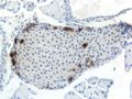
Mouse islet immunostained for pancreatic polypeptide

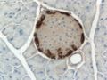
Mouse islet immunostained for insulin
Mouse islet immunostained for glucagon
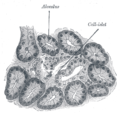
Illustration of dog pancreas. 250x.


انظر أيضا
المصادر
- ^ Langerhans P (1869). "Beitrage zur mikroscopischen anatomie der bauchspeichel druse". Inaugural-dissertation. Berlin: Gustav Lange.
- ^ بسام عبد المسيح. "المعثكلة (غدة ـ)". الموسوعة العربية. Retrieved 2011-12-25.
- ^ Meloche RM (2007). "Transplantation for the treatment of type 1 diabetes". World Journal of Gastroenterology. 13 (47): 6347–55. doi:10.3748/wjg.13.6347. PMID 18081223.
{{cite journal}}: CS1 maint: unflagged free DOI (link) - ^ أ ب Hogan A, Pileggi A, Ricordi C (2008). "Transplantation: current developments and future directions; the future of clinical islet transplantation as a cure for diabetes". Frontiers of Bioscience. 13 (13): 1192–205. doi:10.2741/2755. PMID 17981623.
{{cite journal}}: CS1 maint: multiple names: authors list (link) - ^ Chatenoud L (2008). "Chemical immunosuppression in islet transplantation—friend or foe?". New England Journal of Medicine. 358 (11): 1192–3. doi:10.1056/NEJMcibr0708067. ISSN 0028-4793. PMID 18337609.
- ^ Pileggi A, Cobianchi L, Inverardi L, Ricordi C (2006). "Overcoming the challenges now limiting islet transplantation: a sequential, integrated approach". Annals of the New York Academy of Sciences. 1079 (1): 383–98. doi:10.1196/annals.1375.059. ISSN 0077-8923. PMID 17130583.
{{cite journal}}: CS1 maint: multiple names: authors list (link)
وصلات خارجية
- "THE ISLETS OF LANGERHANS", Karolinska Institutet, Sweden
- "Islets"
- Islet Society
- قالب:MeshNumber
- "Pancreas, human – H&E", Blue Histology – Accessory Digestive Glands, School of Anatomy and Human Biology,