ملف:Muscarinic receptor M2 coupled to protein G - 6OIK.png
حجم هذه المعاينة: 474 × 599 بكسل. البعد الآخر: 1٬700 × 2٬150 بكسل.
الملف الأصلي (1٬700 × 2٬150 بكسل حجم الملف: 2٫47 ميجابايت، نوع MIME: image/png)
وصف قصير
| ⧼wm-license-information-description⧽ |
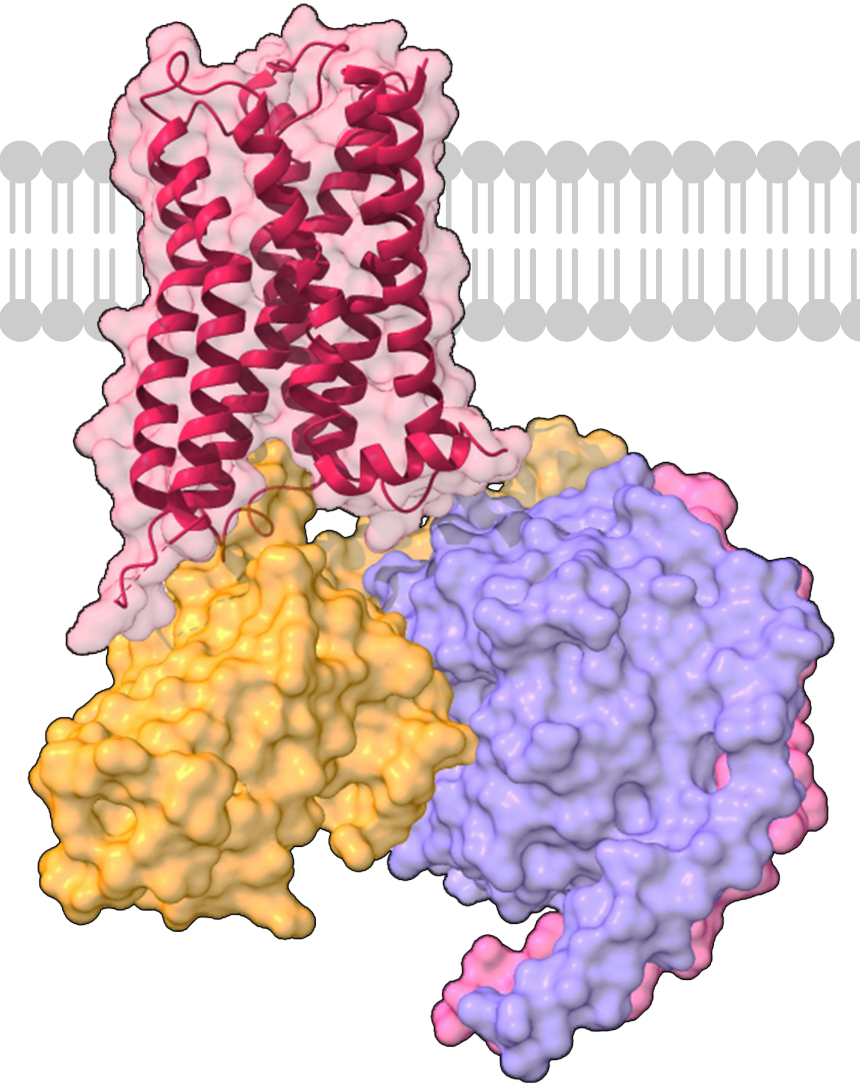
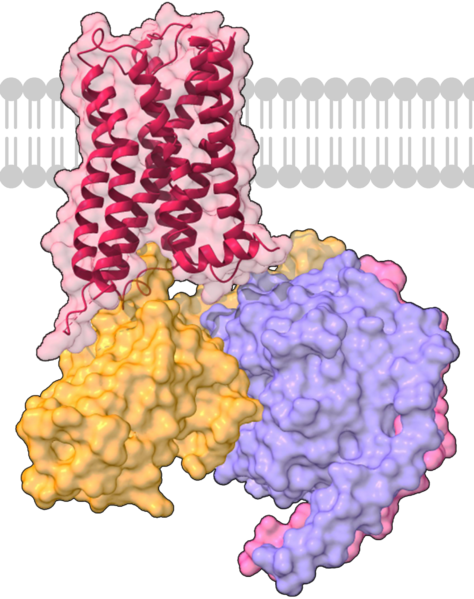
Català: Receptor muscarínic M2 humà acoplat a una proteïna Go. L'estructura tridimensional mostra el receptor M2 (vermell) ancorat a la membrana, la subunitat de proteïna Gαo (taronja) i les subunitats G beta (lila) i gamma (rosa). Imatge creada a partir de l'arxiu PDB 6OIK i representada amb ChimeraX.
Español: Receptor muscarínico M2 humano acoplado a una proteína Go. La estructura tridimensional muestra el receptor M2 (rojo) anclado a la membrana, la subunidad de proteína Gαo (naranja) y las subunidades G beta (lila) y gamma (rosa). Imagen creada a partir del archivo PDB 6OIK y representada con ChimeraX.
English: Human M2 muscarinic receptor in complex with Go protein. The 3D structure shows the M2 receptor (in red) embeded in a membrane, the G protein subunits alpha-o (orange), beta (purple) and gamma (pink). This file is created from PDB file 6OIK and rendered with ChimeraX.
|
| ⧼wm-license-information-date⧽ | 2022 |
| ⧼wm-license-information-source⧽ | ⧼Wm-license-own-work⧽ |
| ⧼wm-license-information-author⧽ | ·Solembum· |
ترخيص
|
تاريخ الملف
اضغط على زمن/تاريخ لرؤية الملف كما بدا في هذا الزمن.
| زمن/تاريخ | صورة مصغرة | الأبعاد | مستخدم | تعليق | |
|---|---|---|---|---|---|
| حالي | ★ مراجعة معتمدة 09:14، 5 نوفمبر 2023 | | 1٬700 × 2٬150 (2٫47 ميجابايت) | Pastakhov (نقاش | مساهمات) | Upload https://upload.wikimedia.org/wikipedia/commons/f/f9/Muscarinic_receptor_M2_coupled_to_protein_G_-_6OIK.png |
لا يمكنك استبدال هذا الملف.
وصلات
لا يوجد صفحات تصل لهذه الصورة.